All about dinosaurs, fossils and prehistoric animals by Everything Dinosaur team members.

Articles, features and information which have slightly more scientific content with an emphasis on palaeontology, such as updates on academic papers, published papers etc.

29 05, 2018

Could We Have Got Pterosaurs All Wrong?

By |2023-10-12T08:01:07+01:00May 29th, 2018|Categories: Dinosaur and Prehistoric Animal News Stories, Dinosaur Fans, Main Page, Palaeontological articles|0 Comments

New Research Challenges Traditional View of Pterosaur Flight

Take a look at a picture of a pterosaur flying and you will see that most illustrations and life-reconstructions of these reptiles depict them travelling through the air with their hind limbs trailing behind them and their back legs wide apart.  However, a new study undertaken by scientists from Brown University (Rhode Island) and the University of California (Berkeley), suggests that we have got this all wrong, pterosaur joints did not permit them to fly with their hind limbs splayed far apart.

Pterosaurs in the Air – But Have we got Their Hind Limbs in the Wrong Position?
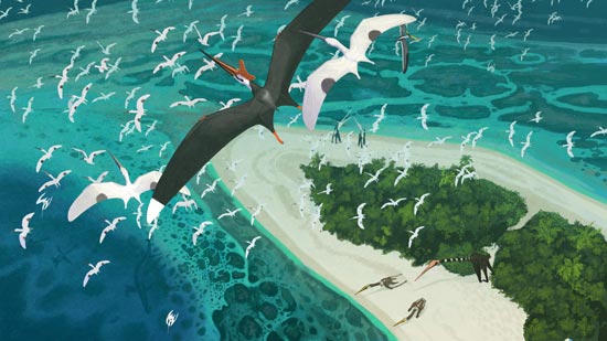
Pterosaurs of the Late Cretaceous (Morocco).
Six new species of pterosaur have been recently identified from Moroccan fossils but if we depict them flying, the convention is to show the hind limbs spread far apart.

Picture credit: John Conway

We Look at the Bones, But What About the Ligaments?

Flying with the hind limbs splayed out, is a posture adopted by most bats when they take to the air.  Ever since the first flying reptiles were described and illustrated, these archosaurs have been depicted in the same way.  However, this new research, published in the Proceedings of the Royal Society B (biology), suggests that ligaments would restrict joint movement and pterosaurs and the volant dinosaurs such as Microraptor, could not have flown in the same way as bats.

Lead author of this study, Armita Manafzadeh, a PhD student at Brown University commented:

“Most of the work that’s being done right now to understand pterosaur flight relies on the assumption that their hips could get into a bat-like pose.  We think future studies should take into account that this pose was likely impossible, which might change our perspective when we consider the evolution of flight in pterosaurs and dinosaurs.”

The “Classical” Pterosaur Flying Posture
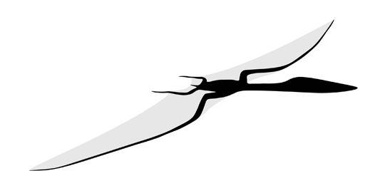
Investigating the flying posture of the Pterosauria.
The “bat-like” posture of the hind limbs of pterosaurs, may not have been anatomically possible.

Picture credit: Armita Manafzadeh

The study undertaken in collaboration with Kevin Padian (University of California), attempted to infer the range of motion of joints in a way that takes into account the soft tissues such as ligaments surrounding the joint.  Usually, soft tissue such as ligament and cartilage does not fossilise, so palaeontologists have to work out joint motion from just the bones alone.  The pair of scientists set out to examine the joint movement of modern dinosaurs – birds, to test the extent to which ligaments influence joint motion.

Chickens at a Grocery Store

Student Manafzadeh explained that the idea started with grocery store chickens:

“If you pick up a raw chicken at the grocery store and move its joints, you’ll reach a point where you will hear a pop.  That’s the ligaments snapping, but if I handed you a chicken skeleton without the ligaments, you might think that its joints could do all kinds of crazy things,  So, the question is, if you were to dig up a fossil chicken, how would you think its joints could move and how wrong would you be?”

Quetzalcoatlus – A Giant Pterosaur Takes to the Air (Note the Splayed Out Back Legs)
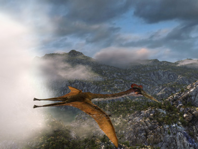
Quetzalcoatlus takes to the air
Quetzalcoatlus takes to the air.

Picture credit: Everything Dinosaur

Dead Quails and X-ray Images

Chickens may be easy to acquire, but for this scientific study, dead quails were used in order to assess joint mobility in a three-dimensional way, rather than just referring to the bones.  Birds are the closest living relatives to the extinct pterosaurs and the non-avian dinosaurs.  Birds (Aves), the Pterosauria and the Dinosauria are members of the Archosauria clade.  This clade is  usually divided into two distinct branches, on one branch (Crurotarsi), are the crocodilians and their ancestors plus several other extinct lineages such as the phytosaurs.  The second branch (Avemetatarsalia) groups all the reptiles more closely related to birds than crocodilians.

A sub-group of the Avemetatarsalia is the Ornithodira, which specifically nests the Pterosauria and their ancestors and the Dinosauria and their ancestors, plus the descendants of dinosaurs – birds, together.  Hence, the use of quail limbs to assess the range of movement and joint mobility.

The skin and muscle surrounding the joints was cut away and once the hip joints were exposed, the scientists manipulated them taking X-ray images to assess the likely range of motion.  By doing this, they could determine the exact positions of the bones in poses where the ligaments restricted and then prevented further joint movement.

Mapping Out the Joint Movements of Ornithodirans

This technique enabled Manafzadeh to map out the range of motion of the quail hip with ligaments attached.  She then compared this range of motion to what was inferred when the bones were considered in isolation.  For the bones-only poses, Manafzadeh used traditional criteria that palaeontologists often employ — stopping where the two bones hit each other and when the movement pulled the thigh bone out of its socket.

This experiment revealed that over 95 percent of the joint positions that seemed plausible with bones alone were actually impossible when the ligaments were attached.

Mapping Out the Range of Motion in Quail Hips
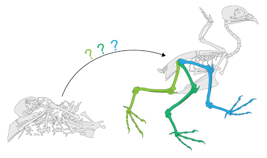
Mapping the movement of Archosaur limbs.
Mapping the range of movement in quail hind limbs to assess the movement of ornithodiran limbs.

Picture credit: Armita Manafzadeh

The Implications for Pterosaurs and the Maniraptora

The team’s next move was to calculate how the range of motion in living birds might correlate to the range of motion expected for extinct pterosaurs and those members of the Maniraptora, such as Microraptor that are believed to have been able to fly.  The assumption has long been that these creatures flew in a similar way to bats.  That is partly because the wings of pterosaurs were made of skin and supported by an elongated fourth finger, which is superficially similar to the wings of bats.  Bat wings are also connected to their hind limbs, which they splay out widely during flight.

Many palaeontologists, Manafzadeh says, assume pterosaurs and four-winged dinosaurs did the same.  But this new study suggests that pose was impossible.

In quail, a bat-like hip pose seemed possible based on bones alone, but outward motion of the thigh bone was inhibited by one particular ligament, a ligament that’s present in a wide variety of birds and other reptiles related to the Pterosauria.  Consequently, in the absence of extraordinary evidence to the contrary, this analysis casts doubt on the “bat-like” hip pose traditionally inferred for pterosaurs and basal maniraptorans and underscores the point that reconstructions of joint mobility based on manipulations of bones alone can be misleading.

To achieve a “bat-like” flying posture, the ligament would have to stretch 63 percent more than the quail ligament can, the implication is that we have been illustrating flying reptiles and flying dinosaurs all wrong.

A Model of a Volant Dinosaur (Microraptor)
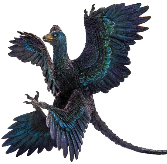
PNSO Gaoyuan the Microraptor model.
The PNSO Gaoyuan the Microraptor model.

The image (above) shows a beautiful Microraptor model from PNSO.

To view this range of prehistoric animal models and figures: PNSO Age of Dinosaurs Models.

In addition to challenging traditional views about flight in pterosaurs and early birds, the research also provides new ways of assessing joint mobility for any joint of any extinct species by looking at its living relatives.

The scientific paper: “ROM Mapping of Ligamentous Constraints on Avian Hip Mobility: Implications for Extinct Ornithodirans” by Armita R. Manafzadeh, Kevin Padian published in the Proceedings of the Royal Society B

Everything Dinosaur acknowledges the assistance of a press release from Brown University in the compilation of this article.

View the award-winning Everything Dinosaur website: Everything Dinosaur.
28 05, 2018

Rare Fossil Flakes and Dinosaur Dandruff

By |2024-05-11T06:01:59+01:00May 28th, 2018|Categories: Dinosaur and Prehistoric Animal News Stories, Dinosaur Fans, Main Page, Palaeontological articles|0 Comments

Dinosaur and Early Bird Dandruff

A team of international scientists led by researchers from University College Cork (Ireland), have discovered the fossilised remains of skin flakes from feathered dinosaurs and primitive birds that lived during the Early Cretaceous.   The fossil flakes of skin, preserved amongst the plumage of the feathered creatures, has provided evidence on how dinosaurs shed their skin.  Unlike many reptiles alive today, animals such as lizards and snakes, which shed their skin as a single piece or as several large pieces, it seems that basal birds and non-avian dinosaurs shed small epidermal flakes just like modern birds and mammals and that includes us with our dandruff.

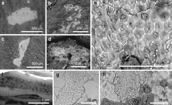
Preserved Soft Tissue Evidence – Flakes of Skin in Maniraptoran Dinosaurs and the Basal Bird Confuciusornis

Fossils of shed dinosaur skin and a basal bird (Confuciusornis).
Phosphatised soft tissues in non-avian maniraptoran dinosaurs and a basal bird.

Picture credit: Nature Communications

Fossil Flakes

The photographs above (labelled a to h), show scanning electron microscope generated images of tissue in the Early Cretaceous Aves C. sanctus (a,e and f), the dromaeosaurid Sinornithosaurus (S. millenii) in photographs c and h, along with the therizinosaur Beipiaosaurus (B. inexpectus), photos b and g.  The photograph labelled d, is a view of epidermal flakes preserved in association with the fossilised remains of Microraptor, which along with Sinornithosaurus is believed to have been capable of flight or at least gliding.

Studying the constituents of flakes of dinosaur skin, which come from animals that might have been volant, is very important.  Palaeontologists can compare these flakes to living birds which are capable of powered flight.  These flakes, in turn can be examined in relation to the flakes of terrestrial feathered dinosaurs such as Beipiaosaurus.  Any differences in the composition of these flakes might provide scientists with further information on the aerial abilities of dinosaurs such as Microraptor and Sinornithosaurus.

An International Team

The scientists include researchers from the Chinese Institute of Vertebrate Palaeontology and Palaeoanthropology, Bristol University, Linyi University (China), the Open University as well as Queen Mary University (London) and the University College Dublin (Ireland).   They studied the fossil cells and dandruff from a range of Early Cretaceous Theropods and compared the skin flakes to those of living, extant birds.

Lead author of the research, Dr Maria McNamara (University College Cork), stated:

“The fossil cells are preserved with incredible detail,  right down to the level of nanoscale keratin fibrils.  What’s remarkable is that the fossil dandruff is almost identical to that in modern birds – even the spiral twisting of individual fibres is still visible.”

Getting to Grips with Corneocytes

The scientists used a scanning electron microscope (SEM), to examine the beautifully preserved, but microscopic fossilised fragments of skin associated with three feathered dinosaur specimens (Microraptor, Beipiaosaurus and Sinornithosaurus) and one Early Cretaceous bird Confuciusornis (Confuciusornis sanctus).  All of these fossils are associated with the Jehol biota of north-eastern China.

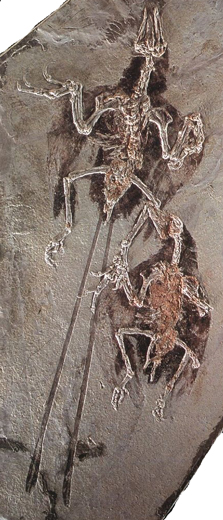
A Pair of Fossilised Confuciusornis (Liaoning Province) Showing the Two Known Body Plans for these Ancient Birds

Confuciusornis fossil birds.
A pair of Confuciusornis fossil birds (Liaoning Province).

Just like our own flakes of skin, our dandruff, the fossil flakes consist of tough cells known as corneocytes.  These cells are full of protein fibres (keratin), such was the quality of preservation that the SEM analysis was able to identify bundles of these fibres and even to hone in on single strands.

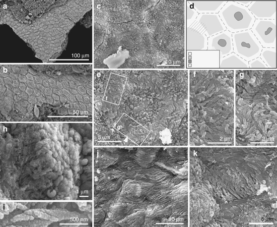
Scanning Electron Micrographs of Skin Flakes Associated with C. sanctus

Fossilised skin flakes of Confuciusornis.
Ultrastructure of the soft tissues in Confuciusornis.

Picture credit: Nature Communications

The photographs (above) show highly magnified images of the skin flakes associated with Confuciusornis.  Closely packed polygons can be observed (a and b), whereas (c) shows a detailed view of the polygons and the first signs at this magnification of the bundles of fibrous keratin.  The drawing (d) interprets the bundles as more darkly shaded areas in the central part of each polygon.  Photograph (e) shows the area that was more closely observed (f and g) indicating the presence of fibres, whereas, (h) shows the fibrous bundle associated with a skin flake in an extreme close-up view.

Helical coiling of the tiny fibres is shown (picture i) and photographs j and k show polygons having been deformed by some form of stretching.

These structures were then compared to the flakes of skin associated with living birds, in this case, male specimens of Zebra Finches (Taeniopygia guttata) and the Java Sparrow (Lonchura oryzivora).  In addition, the fossil evidence was compared to the moulted, downy feathers of a male American Pekin Duck (Anas platyrhynchos domestica).

Fossil Flakes Provide Clues About Dinosaur Metabolism

This research suggests that this ability to constantly shed skin evolved sometime in the late Middle Jurassic, around the same time as a host of other skin features evolved.  The feathered epidermis of dinosaurs acquired many, but not all, anatomically modern attributes close to the base of the Maniraptora clade.

Dr McNamara explained:

“There was a burst of evolution of feathered dinosaurs and birds at this time, and it’s exciting to see evidence that the skin of early birds and dinosaurs was evolving rapidly in response to bearing feathers.”

Co-author, Professor Mike Benton (Bristol University), commented:

“It’s unusual to be able to study the skin of a dinosaur, and the fact this is dandruff proves the dinosaur was not shedding its whole skin like a modern lizard or snake but losing skin fragments from between its feathers.”

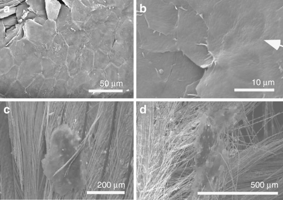
Corneocytes in Living Birds

Corneocytes in living birds
Corneocytes in extant birds.

Picture credit: Nature Communications

The four photographs above, show scanning electron micrographs of shed skin flakes in living birds.  Note the polygonal structure (a), which is reminiscent of the shapes seen when the corneocytes of extinct dinosaurs and birds were studied.  Photograph (b) shows a central depression in the cell associated with a pycnotic nucleus, whilst photographs (c and d) show skin flakes stuck to the bird’s feathers.

Modern birds have very fatty corneocytes with loosely packed keratin, which allows them to cool down quickly when they are flying for extended periods.  The corneocytes in the fossil dinosaurs and birds, however, were packed with tightly bundled keratin, suggesting that the extinct creatures didn’t get as warm as modern birds, presumably because they couldn’t fly at all or for as long.

This suggests that Confuciusornis did not fly that well, if probably flew in short bursts, but may not have been capable of making prolonged flights.  The lack of fatty corneocytes in those dinosaurs which are thought to have had some aerial ability (Microraptor and possibly Sinornithosaurus), sheds doubt on whether they were truly volant.

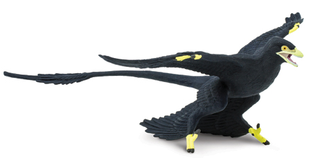
Could Some Dinosaurs Like Microraptor Fly?

Microraptor dinosaur model.
A member of the Dromaeosauridae sub-family the Microraptorinae, but could these dinosaurs fly?  The absence of fatty corneocytes suggests a lower metabolism than with extant birds..

Picture credit: Everything Dinosaur

The picture (above) shows a Microraptor model from Safari Ltd.

To view this range of models and figures: Wild Safari Prehistoric World.

Everything Dinosaur acknowledges the assistance of a press release from the University College Cork (Ireland) in the compilation of this article.

The scientific paper: “Fossilised Skin Reveals Coevolution with Feathers and Metabolism in Feathered Dinosaurs and Early Birds” by Maria E. McNamara, Fucheng Zhang, Stuart L. Kearns, Patrick J. Orr, André Toulouse, Tara Foley, David W. E. Hone, Chris S. Rogers, Michael J. Benton, Diane Johnson, Xing Xu and Zhonghe Zhou published in Nature Communications.

Visit the website of Everything Dinosaur: Everything Dinosaur.

25 05, 2018

How Birds Survived the Cretaceous Mass Extinction Event

By |2023-10-12T08:56:35+01:00May 25th, 2018|Categories: Dinosaur and Prehistoric Animal News Stories, Dinosaur Fans, Main Page, Palaeontological articles|0 Comments

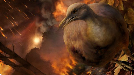
Ground Dwelling Birds Survived Asteroid Strike

One of the fascinating conundrums about the end Cretaceous extinction event is how did the avian dinosaurs (birds) survive, but their very close cousins the non-avian dinosaurs, animals such as Tyrannosaurus rex, Triceratops and Edmontosaurus fail to make it through this calamitous time in Earth’s history?  A team of international researchers, writing in the journal “Current Biology” have put together a fascinating explanation as to why we have birds today, but no other theropods, or indeed any other representatives of the Dinosauria.

With the extra-terrestrial impact event some 66 million and 38 thousand years ago (plus or minus 11,000 years), the ecosystems on our planet were devastated.  Whether this single, huge impact was the sole cause of the mass extinction or whether this was the final “coup de grâce” is debatable, however, life would never be the same again.

Using a variety of data sources, the team, which included scientists from the University of Bath, the Denver Museum of Nature and Science, Yale University and the Field Museum amongst others, have pieced together what the impact event meant for the Aves.  Their scientific paper suggests that the only kinds of birds to survive the Cretaceous-Palaeogene (K-Pg) extinction were ground- dwellers.

How Our Feathered Friends Survived the Cretaceous-Palaeogene Extinction Event

Ground-dwelling birds survived the extinction event.
Ground-dwelling birds survived the K-Pg extinction event.

Picture credit: Phillip Krzeminski

Why Did the Birds Survive?

A number of ideas and theories have been proposed to help explain why the birds are around today, but the non-avian dinosaurs are not.  Recently, Everything Dinosaur published an article on a piece of research that suggested that seed-eating may have contributed to the survival of birds during this devastating time in the history of our planet.

To read the article: Seed Eating May Have Helped the Birds Survive.

Commenting on the scientific paper, lead author Daniel Field of the Milner Centre for Evolution (University of Bath) stated:

“We drew on a variety of approaches to stitch this story together.  We concluded that the devastation of forests in the aftermath of the asteroid impact explains why tree-dwelling birds failed to survive across this extinction event.  The ancestors of modern tree-dwelling birds did not move into the trees until forests had recovered from the extinction-causing asteroid.”

The Collapse of Forests

The scientists analysed the plant fossil record and identified that the world’s woodlands and forests were virtually wiped out by the extra-terrestrial impact.  Huge forest fires would have raged in the immediate aftermath of the impact and it is likely that much of the world had to endure a period of extensive “acid rain” as a result of the catastrophic event.  In the months, or maybe even tens of years afterwards, our planet could have been blanketed in a cloud of dust and ash.  This would have blocked out the sun and led to the collapse of food chains which relied on photosynthesising plants.

The scientists look at the record of fossil pollen and spores to assess the types of flora present and how quickly, ferns, flowering plants (angiosperms) and other types of flora recovered after the extinction event.

Plotting the Turnover of Different Types of Flora from Pollen and Spore Counts

Flora turnover at the K-Pg boundary.
Floral turnover evidenced by changes in relative abundance of common pollen taxa across the K-Pg boundary.

Picture credit: Current Biology with additional annotation by Everything Dinosaur.

The diagram (above), plots the palynological record of the John’s Nose Section in North Dakota, a series of sequential strata laid down before, during and after the extinction event.  It helps to plot the demise of different types of plant and their recovery (floral turnover), as evidenced by changes in relative abundance of common pollen taxa across the K-Pg boundary.  Note, the “fern spike” recorded not long after the extinction event, ferns are usually the first type of plant to recover from natural disasters today, as evidenced by their ability to re-populate areas destroyed following volcanic activity.

Evolutionary Relationships of Living Birds

The research team examined the evolutionary relationships of extant birds and their ecological habits to map how bird ecology has changed over time.  The data revealed that the most common ancestor of all living birds, all the bird lineages that survived the K-Pg extinction event, most likely were ground-dwellers.  In contrast, many Aves that lived at the end of the age of dinosaurs (and there were lots of them), exhibited tree-dwelling, arboreal habits.  These species did not survive the mass extinction event and therefore they have no direct living descendants around today.

Daniel Field added:

“Today, birds are the most diverse and globally widespread group of terrestrial vertebrates, there are nearly 11,000 living species.  Only a handful of ancestral bird lineages succeeded in surviving the K-PG mass extinction event 66 million years ago and all of today’s amazing living bird diversity can be traced to these ancient survivors.”

The researchers conclude that their findings shed light on the fundamental influence major events in the history of our planet have on the evolution of living things.  The team hope to build on this initial research and to explore the timing of the recovery of the vegetation and to develop a better understanding of the early radiation of the birds.  After all, those lucky survivors inherited a brave new world, devoid of non-avian dinosaurs and many other terrestrial and marine organisms too.

The scientific paper: “Early Evolution of Modern Birds Structured by Global Forest Collapse at the End-Cretaceous Mass Extinction” by Daniel J. Field, Antoine Bercovici, Jacob S. Berv, Regan Dunn, David E. Fastovsky, Tyler R. Lyson, Vivi Vajda and Jacques A. Gauthier published in the journal Current Biology.

Visit the Everything Dinosaur website: Everything Dinosaur.
11 05, 2018

A New New Zealand Pigeon from the Miocene

By |2023-10-13T07:47:31+01:00May 11th, 2018|Categories: Dinosaur and Prehistoric Animal News Stories, Main Page, Palaeontological articles|0 Comments

The Zealandian Dove Related to the Dodo

Scientists from the Canterbury Museum (New Zealand), in collaboration with colleagues from the University of New South Wales, Flinders University and the Museum of New Zealand Te Papa Tongarewa, have described a new species of prehistoric pigeon that flapped around South Island during the Miocene Epoch, some 16-19 million years ago.  The new species has been named the Zealandian Dove (Deliaphaps zealandiensis) and it may have been related to the extinct, giant, flightless pigeon of Mauritius – the Dodo.

Part of the Dodo Collection from the Canterbury Museum 

Canterbury Museum Dodo exhibit.
Casts of the Oxford Museum Dodo specimen which is part of the Canterbury Museum collection.

Picture credit: Canterbury Museum

New Zealand only has two species of native pigeons, the aptly named New Zealand pigeon, otherwise called the Kereru (Hemiphaga novaeseelandiae) and the closely-related Chatham Island pigeon, or Parea (Hemiphaga chathamensis).

The fossils were found at a dig site near St Bathans (Central Otago, South Island) and although fragmentary in nature, consisting of a few wing bones and part of the pectoral girdle, the researchers are confident that this material represents a new species of prehistoric pigeon and have published their paper in the “Paleontología Y Evolución de las Aves”.

The Zealandian Dove

Deliaphaps zealandiensis has been named after the landmass called Zealandia.  This large area of land is also referred to as the New Zealand continent or Tasmantis.  It consists of a mass of continental crust that sank after breaking away from Australia 60–85 million years ago, having begun to separate from Antarctica and the rest of the Gondwana supercontinent.  Most of Zealandia is submerged, but it rises above water in places, including the populated areas of New Zealand, New Caledonia, Norfolk and the Lord Howe Island group.

Sixteen Years to Gather the Fossil Material

The fossil material has been slowly gathered over sixteen years and one of the wing bones is similar to the wing bones of members of a group (the Raphinae), that includes the Tooth-billed pigeon (Didunculus strigirostris) or Manumea which is native to the island of Samoa.  The Raphinae also includes the Crowned pigeons of New Guinea (Goura genus) and the spectacular Nicobar pigeon of south-east Asia (Caloenas nicobarica), believed to be the closest living relative of the enigmatic Dodo.

The Newly Described Zealandian Dove is Probably Closely Related to the Nicobar Pigeon

The Nicobar pigeon.
The Nicobar pigeon, the closest living relative to the Dodo. It may also be related to the newly described Zealandian Dove from the Miocene.

Picture credit: Canterbury Museum

Co-author of the scientific paper and University of New South Wales scientist, Professor Sue Hand stated:

“Fossils recovered from the St Bathans site now number in the thousands and together document a time of great biodiversity in New Zealand’s history.  For many of New Zealand’s very distinctive bird lineages, such as moa and kiwi, the St Bathans fossils provide their oldest and sometimes first deep time records.  Discovery of the Zealandian Dove and its evident links to the dodo are fascinating additions to the unfolding picture of New Zealand’s prehistoric menagerie.”

A Miocene Pigeon

Lead author of the research, Dr Vanesa De Pietri (Canterbury Museum), added:

“Based on the St Bathans fossils, we think that the Zealandian Dove is part of this Indo-Pacific group.  It is probably most similar to the Nicobar Pigeon and is therefore a close relative (or at least a cousin) of the famous dodo.  The Zealandian Dove is the first record of this group found in the southern part of the nearly submerged land mass known as Zealandia.”

Second Pigeon from St Bathans (Miocene Deposits)

The Zealandian Dove is only the second pigeon found at the St Bathans fossil site.  The delicate and fragile bones of birds are not strong candidates to endure the fossilisation process.  The first pigeon to be named from fossils discovered at this location was the St Bathans pigeon (Rupephaps taketake), which was described from a single coracoid bone recovered from these ancient lake deposits.  It is believed to be related to the extant, native New Zealand pigeons.

The Oxford Museum Dodo Material

Oxford Museum Dodo.
The Oxford Museum Dodo material.

Picture credit: Oxford Museum

Commenting on the recent pigeon fossil discoveries, Dr Paul Scofield of Canterbury Museum said:

“We have now also found a leg bone that we can attribute to this species [St Bathans pigeon].  As a result, we can now confirm that the St Bathans pigeon is also closely related to Indonesian and Melanesian mountain pigeons.  It was an early offshoot within that particular group.”

Dr Trevor Worthy of Flinders University (South Australia) added:

“Some 19 to 16 million years ago, the diversity of endemic pigeons in New Zealand included at least two distinct co-existing lineages in the southern part of Zealandia taking advantage of the more diverse fruiting trees then available.  Pigeon fossils are rare in the St Bathans fauna and are outnumbered by about thirty to one by parrots, which perhaps reflects the relative abundance of these tree-dwelling birds in the St Bathans fauna.  Many small parrots form large flocks, whereas pigeons typically live in only small groups, so perhaps these traits typified the early Miocene parrots and pigeons in Zealandia.”

Evidence of Climate Change

The reduction in the number of species found in New Zealand is probably a result of climate change which affected the types of trees on the islands.  Scientists know that between approximately 14.2 and 13.8 million years ago, a period of dramatic global climatic cooling took place.  Prior to the global cooling, New Zealand enjoyed a subtropical climate which led to a very diverse and rich fauna and flora with many kinds of fruit-bearing trees that would have provided food for the pigeons.  The loss of the floral diversity as a result of climate change would have had a big impact on the fruit and seed-eating birds and this could be a reason for the reduction in pigeon species.

Everything Dinosaur acknowledges the assistance of a media release from Canterbury Museum in the compilation of this article.

The Everything Dinosaur website: Everything Dinosaur.
9 05, 2018

A Remarkable Weather Forecast from the Cambrian

By |2024-05-10T19:04:08+01:00May 9th, 2018|Categories: Dinosaur and Prehistoric Animal News Stories, Geology, Main Page, Palaeontological articles, Photos/Pictures of Fossils|0 Comments

Tiny Fossils Provide Clues to Earth’s Climate 500 Million Years Ago

A joint team of scientists from France and the UK, have plotted the temperature of our planet’s oceans over half a billion years ago using a combination of fossil data and computer-based climate models.  Think of it as a sort of weather forecast from the Cambrian.

A Cambrian Climate Study

This newly published research suggests that the first hard-bodied animals diversified in warms seas, heated by a greenhouse world.  The team’s findings help to expand our knowledge of the environment at the time of the Cambrian explosion, a period in Earth’s history that saw a huge increase in the number and type of marine animal forms.

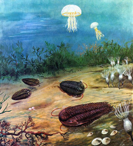
Life in the Late Cambrian Period

Cambrian life.
Life in the Late Cambrian by Zdeněk Burian.

Picture credit: Zdeněk Burian

Writing in the academic journal “Science Advances”, the scientists, led by researchers from the University of Leicester, used climate models and the chemical analysis of tiny, shelly fossils preserved in limestone from Shropshire (central England), to calculate the sea temperature during a time of rapid diversity of animal life in the Palaeozoic.

From around 540 to 510 million years ago, the fossil record shows a marked change, as during this period of Earth’s history, virtually all of the animal phyla (including the Chordata – our phylum) appeared.  The idea of a “Cambrian explosion” is a little misleading, the appearance of many new forms of complex animal life may have been gradual, but in terms of the fossil record, sites such as the famous Burgess Shale of British Columbia and Yunnan Province (southern China), have revealed extensive and varied marine ecosystems with large numbers of new types of animal being recorded in the strata.

Analysis of Some of the First Shelly Fossils

Scientists had thought that for much of the Cambrian, our planet was warmer that it is today with no polar ice caps present.  A study of tiny 1 mm long fossils of some of the first animals to produce a hard, shelly exoskeleton has confirmed this hypothesis.  Analysis of isotopes from the tiny shells in combination with the climate models show that at high latitudes (around 65 degrees south), sea temperatures were in excess of 20 degrees Celsius.

This might seem very warm, especially when you consider that this is an evaluation of sea temperatures at approximately 65 degrees south, today, travelling to that latitude would put you on the southernmost fringes of the Southern Ocean and close to Antarctica.  However, the data generated is similar to more recent, better understood, greenhouse climates such as that of the Late Cretaceous.

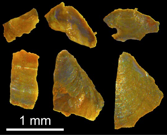
Reflected Light Microscopy – Brachiopod Fossils Used in the Study

Reflected light microscope images of Cambrian brachiopods.
Reflected light microscope images of some of the brachiopod fossils (phosphatic microfossils), used in this study.

Picture credit: Leicester University

Analysing Isotopes

Co-author of the open access paper, PhD student Thomas Hearing (University of Leicester’s School of Geography, Geology and the Environment), explained:

“Because scientists cannot directly measure sea temperatures from half a billion years ago, they have to use proxy data, these are measurable quantities that respond in a predictable way to changing climate variables like temperature.  In this study, we used oxygen isotope ratios, which is a commonly used palaeothermometer.  We then used acid to extract fossils about 1 mm long from blocks of limestone from Shropshire, UK, dated to between 515 – 510 million years old.  Careful examination of these tiny fossils revealed that some of them have exceptionally well-preserved shell chemistry which has not changed since they grew on the Cambrian sea floor.” 

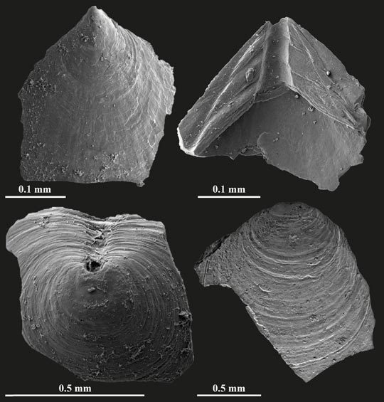
High Resolution Scanning Electron Microscope (SEM) Images of Brachiopod Fossils Used in the Study

SEM images of brachiopods.
Electron microscope images of some of the brachiopod fossils used in this study. Electron microscopy allows much higher resolution imaging of small structures than normal light microscopy.

Picture credit: Leicester University

For models and replicas of invertebrates, including trilobites and other Cambrian animals: CollectA Prehistoric Life Models and Figures.

Identifying Chemical Signatures in Cambrian Fossils

Dr Tom Harvey (University of Leicester) added:

“Many marine animals incorporate chemical traces of seawater into their shells as they grow.  That chemical signature is often lost over geological time, so it’s remarkable that we can identify it in such ancient fossils.” 

Analyses of the oxygen isotopes of these fossils suggested very warm temperatures for high latitude seas (~65 °S), probably between 20 °C to 25 °C.  To see if these were feasible sea temperatures, the researchers carried out climate model simulations for the Cambrian.  The climate model scenarios suggest that the Earth’s climate was in a “typical” greenhouse state, with temperatures similar to more recent and better understood greenhouse intervals known from the Mesozoic and the Cenozoic eras.  Ultimately, this study will help to expand our knowledge of the ecosystem that existed during the Cambrian.

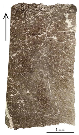
The Highly Fossiliferous Comley Limestones (Shropshire, UK)

A thin section of highly fossiliferous rock of Cambrian age.
A thin section slice through the trilobite-rich Comley Limestones (Shropshire, UK).

The curves and white wavy lines in the photograph (above), are preserved exoskeletons of numerous trilobites.

Thomas Hearing concluded:

“We hope that this approach can be used by other researchers to build up a clearer picture of ancient climates where conventional climate proxy data are not available.”

The research was carried out as an international collaboration involving scientists from the University of Leicester (UK), British Geological Survey (BGS; UK), and CEREGE (France).

Everything Dinosaur acknowledges the help of Leicester University in the compilation of this article.

Visit the Everything Dinosaur website: Everything Dinosaur.
6 05, 2018

Dense Bones and Other Aquatic Adaptations in Spinosaurs

By |2023-10-13T09:03:04+01:00May 6th, 2018|Categories: Dinosaur and Prehistoric Animal News Stories, Dinosaur Fans, Main Page, Palaeontological articles|1 Comment

South American Giant Provides Further Information on Enigmatic Spinosaurs

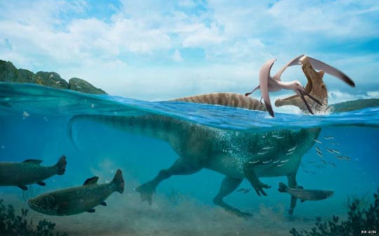
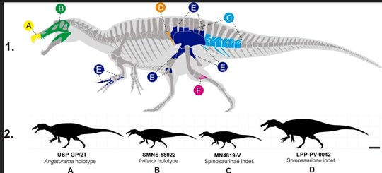
A partial leg bone found in north-eastern Brazil has helped scientists to better understand the adaptations members of the Spinosauridae may have evolved to help them with their semi-aquatic lifestyles.  Furthermore, the fragmentary fossil, a large partial tibia from the Aptian-Albian Romualdo Formation, (Araripe Basin, north-eastern Brazil), when compared to other spinosaur remains, indicates an individual dinosaur much larger than other South American spinosaurids.  This single fossil suggests a sub-adult animal around ten metres in length, far larger than the other South American spinosaurids from the Araripe Basin such as Irritator and Angaturama.

Studying the Spinosauridae

A South American Lagoon Around 115-110 Million Years Ago – A Spinosaurid Attacks a Pterosaur

Spinosaur attacks a Pterosaur.
An illustration of a South American Spinosaur attacking a pterosaur.

Picture credit: Julio Lacerda

For models and replicas of members of the Spinosauridae and other theropod dinosaurs: CollectA Deluxe Prehistoric Life Models.

Sail-back Dinosaur from the Heart of the Brazilian Outback

The Brazilian dinosaur fossil is providing another piece of the puzzle as palaeontologists strive to better understand the enigmatic spinosaurids.  The discovery of the fossil bone and its implications for Spinosaur research has been published in the academic journal “Cretaceous Research”.  The research was led by a team of Brazilian palaeontologists in collaboration with colleagues from Yale University, the University of Bonn and Trinity College (Dublin).

The partial tibia (lower leg bone) was found in Ceará, a state in the heart of the Brazilian outback.  Although only a fragment of bone, its size in relation to other spinosaurid fossils suggests a dinosaur measuring about ten metres in length, considerably bigger than other South American members of the Spinosauridae.

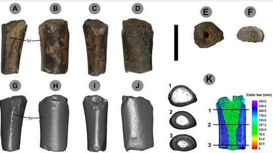
The Partial Tibia Bone (Various Views)

Views of the Spinosaur fossil material
Views and cross-sectional analysis of the spinosaur partial tibia fossil (LVP-PV-0042).

Picture credit: Cretaceous Research

Large Predators with an Aquatic Lifestyle

Over the last five years or so, there have been a number of papers published looking at how these large predators lived.  Many palaeontologists believe that these theropods adopted a specialist lifestyle, becoming semi-aquatic and essentially piscivores.  Interest in these enigmatic dinosaurs has certainly been piqued in recent years, especially with the publication of a fascinating paper in 2014 that proposed that Spinosaurus aegyptiacus was a semi-aquatic, obligate quadruped.

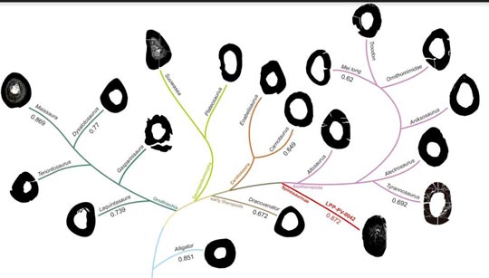
The partial tibia has anatomical traits previously only observed in the north African S. aegyptiacus, traits such as a reduced fibular crest and dense bones (osteosclerotic bones).  These types of bones are characteristic of the limb bones of vertebrates that spend a lot of their time in water.  The leg bones of hippos, for example, exhibit this condition.  The partial tibia from north-eastern Brazil, supports the idea that spinosaurids were adapted to an aquatic environment, in addition, the Brazilian fossil is many millions of years older than those fossils associated with S. aegyptiacus, so, this suggests that high bone compactness was already present in Brazilian spinosaurids long before S. aegyptiacus evolved.

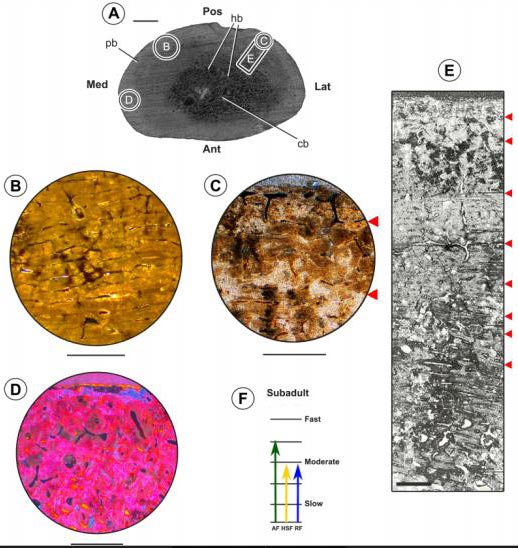
Cross-sectional Views of LPP-PV-0042 Indicating Bone Density and Growth Rate

Growth rate and density of Araripe Basin dinosaur bone.
An assessment of the bone density observed in LPP-PV-0042 along with histology of bone indicating growth rate.

Picture credit: Cretaceous Research

Fossilised bone cells in detail, observed under the microscope.  Different growth pulses of the animal are represented by the red arrows.  Absence of certain characteristics in the bone tissue lead palaeontologists to conclude the spinosaurid was a sub-adult and still growing when it died.

To read Everything Dinosaur’s article on the 2014 Spinosaurus paper: Spinosaurus – Four Legs are Better Than Two.

“Heavy Bones” of the Spinosauridae

The study was led by postgraduate student Tito Aureliano (Campinas State University, Unicamp, Brazil), along with researchers from the Federal University of Sao Carlos (Brazil).  The dense bones of the Brazilian Spinosaur would have helped the animal to dive and to move in water in a similar way to a hippopotamus.

The dense bones are analogous to the lead weights used by divers to counteract their own buoyancy.  This research suggests that osteosclerotic bones were present in the South American Spinosaurinae at least ten million years earlier than those associated with their north African cousins.  It is not known when osteosclerotic bones evolved in spinosaurs, but this characteristic may have evolved relatively early in these types of dinosaurs.

Bone Density Comparisons

Spinosaur limb bones are much more dense than most other dinosaurs
Cross sections of vertebrate bones including many dinosaurs showing bone density versus pneumaticity within the Dinosauria.

Picture credit: Cretaceous Research

Commenting on the significance of this study, Tito Aureliano stated:

“It may be possible that Brazilian spinosaurs were the first to adopt this way of life.  Now we need to investigate an even older species.”

An Example of Convergent Evolution

Several types of not-closely related vertebrates have dense bones, reflecting an adaptation to an aquatic lifestyle.  Penguins, crocodiles and sealions along with hippos and spinosaurids have these types of bones, this is an example of convergent evolution.

Aline Ghilardi (Federal University of Sao Carlos) explained:

“It is interesting how this adaptation evolved multiple times in different groups of animals that adopted the same lifestyle.”

The spinosaurids evolved in a different direction when compared to most of the Theropoda.  Whilst most dinosaurs evolved ways to make the skeleton lighter, epitomised in the extreme pneumaticity observed in birds, the spinosaurs developed a way to make their skeleton heavier.  This aided them when it came to occupying a very distinct and specialised niche amongst the Dinosauria.

South American Giants

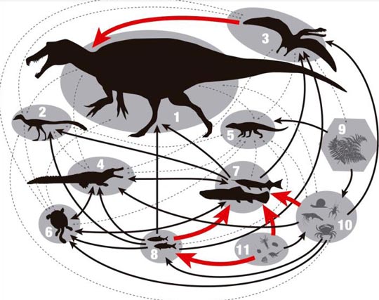
South America might be famous for its huge Cretaceous plant-eating dinosaurs such as the titanosaurids Argentinosaurus, Patagotitan and Dreadnoughtus but the partial tibia bone hints at super-sized predators too.  An analysis of the fossilised cells showed that the studied dinosaur had not reached its maximum size by the time of its death and was still growing.  This indicates that these dinosaurs could reach larger sizes than previously thought, the Araripe Basin Spinosaurinae could have been giants, which would make them the top predators of the Cretaceous coastal lagoons of Ceará.

South American Spinosaur Size Comparison

Spinosaur size comparison from the Araripe Basin.
Spinosaurid size comparison based on known fossil material.

Picture credit: Cretaceous Research

The image (above), shows all the known spinosaurid fossils from the Araripe Basin.  The fossil used in this study is marked in pink (F).   Note, figure 1 shows the fossil bones not to scale but figure 2 provides a scale comparison between the spinosaur specimens from the Araripe Basin.  The largest spinosaurid found to date (LPP-PV-0042), was the subject of the research paper.

Pterosaur Vertebrae from Ceará

The Ceará state has been synonymous with amazing spinosaur fossil finds.  In 2004, researchers led by the famous French palaeontologist Eric Buffetaut, described a remarkable fossil find from this region, three articulated pterosaur vertebrae (Ornithocheiridae) were found with the tooth of a spinosaurid embedded in one of the bones.  This fossil represents direct evidence that spinosaurs included other prey items in their diets as well as fish.  The broken tooth also provides evidence that the light bones of pterosaurs were much stronger than previously assumed.

A Food Chain of the Lagoonal Environment (Araripe Basin in the Early Cretaceous)

Araripe Basin (lagoon) food chain.
Proposed food chain showing LPP-PV-0042 as apex predator.  pterosaurs were on the menu to.

Picture credit: Cretaceous Research

Lots of questions about the Spinosauridae remain.  For example, whether these aquatic adaptations are related to the establishment of a large system of lagoons between South America and Africa caused by the opening of the Atlantic Ocean during this period in Earth’s history.  As with many whales today, was the adaptation of an aquatic lifestyle the trigger that enabled these theropods to evolve into giants?

Our thanks to Tito Aureliano ((Campinas State University, Unicamp, Brazil) for their help in the compilation of this article.

The scientific paper: “Semi-aquatic Adaptations in a Spinosaur from the Lower Cretaceous of Brazil” by Tito Aureliano, Aline M. Ghilardi, Pedro V. Buck, Matteo Fabbri, Adun Samathi, Rafael Delcourt, Marcelo A. Fernandes and Martin Sander published in the journal Cretaceous Research.

Visit the Everything Dinosaur website: Everything Dinosaur.
3 05, 2018

The First Beak Under the Noses of Scientists

By |2023-10-13T21:10:49+01:00May 3rd, 2018|Categories: Dinosaur and Prehistoric Animal News Stories, Dinosaur Fans, Main Page, Palaeontological articles, Photos/Pictures of Fossils|0 Comments

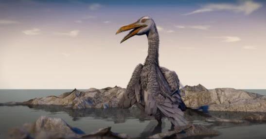
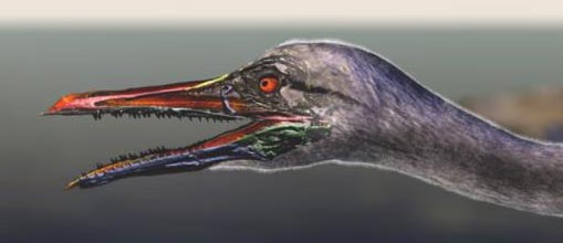
Inspiring Ichthyornis – Top of the Pecking Order

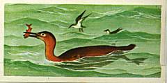
As a very young boy, I remember eagerly striving to complete my Brooke Bond “Prehistoric Animals” card collection.  This was a set of fifty cards to collect,  given away free with packets of tea.   One of the cards featured a pair of toothed, prehistoric birds, a large, reddish coloured Hesperornis which was being mobbed by a couple of tern-like birds, this was my first introduction to Ichthyornis.  Perhaps, the first time that I realised that birds (at least primitive, toothed birds), lived alongside dinosaurs.

How wonderful to read this week that Ichthyornis, thanks to a pieced together three-dimensional skull, may be providing palaeontologists with fresh insights into avian evolution.  The Hesperornis/Ichthyornis picture card may have been burned into my conscience long ago, but it is refreshing to think that this ancient bird may represent a pivotal moment in the transition from dinosaurs to modern-day birds and its significance has only just come to light.  A team of international scientists have published a paper proposing that Ichthyornis may have had one of the first, true bird-like beaks.

The Brooke Bond Picture Card

Hesperornis and Ichthyornis
Hesperornis catching a fish, with Ichthyornis in close attention.

Picture credit: Everything Dinosaur/Brooke Bond

Toothy Bird with the Beginnings of a Beak

Writing in the journal “Nature”, researchers report on the analysis of beautifully preserved three-dimensional Ichthyornis (I. dispar) fossil skull that is providing new evidence on the evolution of the avian head and how the skull and beaks of birds evolved from their dinosaurian ancestors.

A Three-Dimensional Image of Ichthyornis Skull Material Indicates the Tip of the Premaxillary Formed the First Beak

The tip of the premaxillary forms the first beak.
A computer generated image showing the life position of the fossil bones in the three-dimensional Ichthyornis skull.

Picture credit: Yale Office of Public Affairs and Communications

Ichthyornis dispar

Known from fragmentary fossils from Kansas and named back in 1872 by Yale University’s Othniel Charles Marsh, it seems fitting that this new study into one of the first toothed birds described, has been led by scientists from Yale University.  Working in conjunction with colleagues from the University of Kansas, Fort Hays State University, Alabama Museum of Natural History and the McWane Science Centre (Alabama), the team report on new specimens with three-dimensional cranial remains, including one example of a complete skull and two previously overlooked cranial elements that were part of the original Yale specimen examined by Marsh.

Using CT scans and sophisticated computer modelling, individual skull and jaw bones were scanned and reproduced in three-dimensions.  This allowed a complete skull to be constructed revealing new details about the transition from dinosaur skull to a more modern bird skull.

The Transition from Dinosaur Skull to Bird Skull

Yale University palaeontologist and lead author of the study Bhart-Anjan Bhullar commented:

“Right under our noses this whole time was an amazing, transitional bird.  It has a modern-looking brain along with a remarkably dinosaurian jaw muscle configuration.”

Ichthyornis is part of the biota of the Western Interior Seaway, a shallow sea that split North America in two during the Late Cretaceous.  It has been regarded as an early version of a tern or gull, but its size is unknown as the few fossils found represent individuals of different sizes, however, it probably had a wingspan of no more than sixty centimetres, making Ichthyornis slightly smaller than today’s Common Tern (Sterna hirundo), a bird which fills the same ecological niche as the Mesozoic Ichthyornis.

Using the Latest Research, a New Reconstruction of Ichthyornis dispar was Produced

Ichthyornis life reconstruction.
A life reconstruction of Ichthyornis.

Picture credit: Yale Office of Public Affairs and Communications

Ichthyornis – The Evolution of a Beak

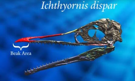
Having built a three-dimensional model of the skull and jaw bones, the researchers were able to note that the premaxillary bone in the upper jaw had become elongated and this, working in conjunction with a keratinous tip on the lower jaw formed the first “proto-beak”.  Ichthyornis dispar shows scientists what the first type of bird beak looked like.  This beak may have evolved as the function of the hands was increasingly limited as they were adapted to form a more effective wing.

The grasping hands of the maniraptoran dinosaurs were no longer able to grasp and manipulate objects so the jaws had to take on an additional function, secondary to their main function – dispatching and consuming prey.

The Beak of Ichthyornis

The beak of Ichthyornis.
The beak of Ichthyornis evolving to replace grasping, functional hands and fingers.

Picture credit: Yale Office of Public Affairs and Communications

Although maniraptoran dinosaurs may not have been able to pronate their hands like us and they lacked an opposable thumb, as forelimbs and hands evolved into wings, so the jaws took over the function of the digits and manus.

Bhart-Anjan Bhullar stated:

“The first beak was a horn-covered pincer tip at the end of the jaw.  The remainder of the jaw was filled with teeth.  At its origin, the beak was a precision grasping mechanism that served as a surrogate hand as the hands transformed into wings.”

The research team conducted its analysis using CT-scan technology, combined with specimens from the Yale Peabody Museum of Natural History; the Sternberg Museum of Natural History in Hays, Kansas, the Alabama Museum of Natural History; the University of Kansas Biodiversity Institute and the Black Hills Institute of Geological Research (South Dakota).

Bird Beaks versus Bird-hipped Dinosaur Beaks

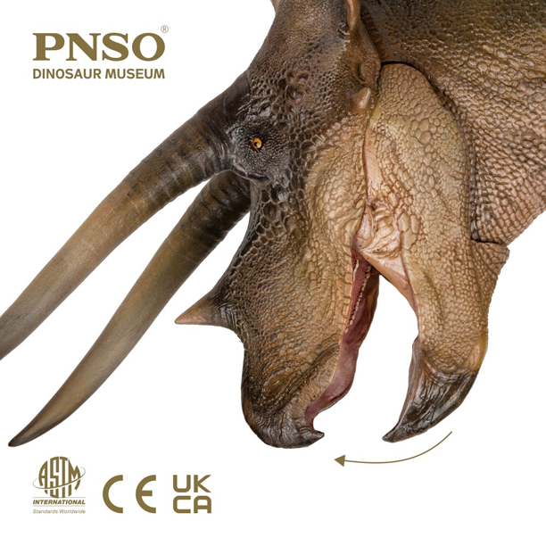
The modern bird beak is a unique organ amongst vertebrates, although notably most derived ornithischian (bird-hipped) dinosaurs possessed a beak, formed from the unique predentary bone in the lower jaw and a roughened, extension of the premaxilla (or the rostral in the case of ceratopsians), in the upper jaw, which allowed the attachment of a keratinous tip which in conjunction formed the beak-like structure – believed to be an adaptation to assist with cropping vegetation.

This study of Ichthyornis suggests that the first bird beak was not the long organ seen in modern birds, but a little pincer tip to grasp and manipulate objects.

A Chasmosaurine Ceratopsian with the Roughened Rostral and the Predentary Forming a Plant-cropping Beak

PNSO Doyle the Triceratops model has an articlated lower jaw
The PNSO Doyle the Triceratops model has an articulated jaw.

The picture (above) shows a ceratopsian figure from the PNSO model range.

To view these figures: PNSO Age of Dinosaurs Models.

Fresh Insight into the Evolution of Extant Bird Skulls

The scientists conclude that their study offers new insights into how modern birds’ skulls formed.  Along with its transitional beak, Ichthyornis dispar had a brain similar to that seen in extant birds but a temporal region of the skull that was reminiscent of a dinosaur.  This suggests that during the evolution of Aves, the brain transformed first, possibly to adapt to a volant (aerial) lifestyle, whilst the remainder of the skull retained the ancestral features associated with the Dinosauria.

Ichthyornis retained a large adductor chamber bounded at the top by substantial bony remnants of the ancestral reptilian upper temporal fenestra (hole in the skull).  This combination of features indicates that important attributes of the avian brain and palate evolved before the reduction of jaw musculature and the full transformation of the beak.

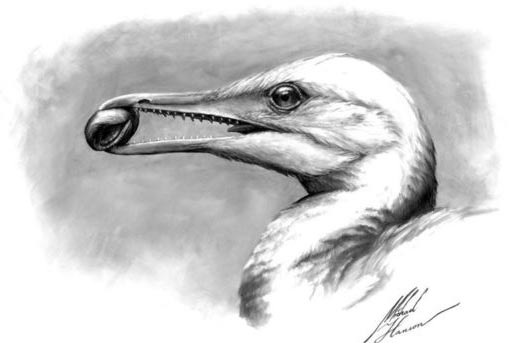
The Beak of Ichthyornis Grasping a Mollusc

Holding a mollusc in its beak.
An illustration of an Ichthyornis holding a mollusc in its beak.

Picture credit: Michael Hanson/Bhart-Anjan Bhullar

I may never have completed my Brooke Bond card collection, but at least, thanks to this new Ichthyornis study, our understanding of the evolution of the beak in birds is more complete.

Visit the Everything Dinosaur website: Everything Dinosaur.
1 05, 2018

Unlocking the Secrets of the Insect in Amber Fossilisation Process

By |2023-10-13T21:23:38+01:00May 1st, 2018|Categories: Dinosaur and Prehistoric Animal News Stories, Main Page, Palaeontological articles|1 Comment

Preservation Bias in Amber Fossilisation Examined

Fossils preserved in amber can give scientists unique insights into ancient ecosystems.  Petrified tree resin can provide a record of some of the smaller members of a prehistoric woodland habitat such as the insects, mites and spiders.  Spores and pollen grains trapped inside a nodule allow palaeobotanists the opportunity to assess the composition of the flora of a 100-million-year-old Cretaceous tropical forest, but all may not be as it seems.  Fossils in amber appear, on their surface, to be perfectly preserved, which in turn suggests that the amber fossil record is perfect and unbiased.  This is not the case, all that glitters inside an amber nodule may not be hidden palaeontological treasure.

A Mosquito Preserved in Amber – But How Much of the Insect is There?

Mosquito fossil preserved in amber.
Amber can provide a window into ancient ecosystems, but what factors affect the preservation process?

Picture credit: Oregon State University

Fossils Preserved in Amber

However, recent advances in three-dimensional imaging techniques, specifically synchrotron tomography, have allowed researchers to look inside amber-entombed fossils and observe that preservation is highly variable.  Many specimens are lacking some or all internal soft tissues, and some specimens are even lacking the more decay-resistant cuticle and are simply empty moulds stained in a life-like colour by remnant organic carbon.

With so many amazing discoveries being made, particularly in burmite from Myanmar, a team of scientists set about examining the amber fossil record in order to assess any potential preservation bias that might exist, with a focus on prehistoric insects.

In the project, the researchers used a serious of laboratory experiments to test the effect of three variables, resin (the un-fossilised precursor to amber) chemistry, gut biota, and dehydration prior to entombment, on the decay of a fruit fly engulfed in resin to better understand the controls on the fossil record of insects in amber.

The team discovered that resin chemistry has a large effect on decay: flies entombed in Wollemia (W. nobilis) tree resin retained essentially all of their external and internal morphology even after one and a half years, whereas flies entombed in Pinus (Scots Pine P. sylvestris) tree resin, were nothing but empty moulds after the same length of time.

Fruit Flies Being Entombed in Tree Resin

Fruit flies (arrowed) become trapped in tree resin.
Part of the experiment – fruit flies being embedded in tree resin.  The fruit flies are indicated by the arrows.

Gut biota had a smaller effect on decay: flies with an intact gut microbiota showed more rapid decay, as indicated by more extensive production of decay gases, than flies that were treated with an antibiotic prior to entombment.  Dehydration prior to entombment also enhanced decay, presumably because resin has very effective decay-inhibiting properties, and therefore any delay in embedding a carcass in resin enhances decay.  These three variables influence the preservation of fossils in amber, and therefore can impart a bias on the fossil record of insects in amber.  Writing in the academic journal “PLOS One”, the researchers concluded that, in particular, resin chemistry and gut biota may strongly influence the amber fossil record.

Synchrotron Images and a Three-Dimensional Reconstruction of the Fruit Fly Being Constructed

A synchrotron was used to scan the fruit flies entombed in the tree resin.
A synchrotron scan of the experiments, showing three different planes of the scan, and a 3-D reconstruction in progress.

The Chemistry of Tree Resin

Resin chemistry is the most likely control on whether or not a specific fossil site preserves the most decay-prone morphological features on the most decay-prone components of an ecosystem.  This could be a particularly confounding bias in the amber fossil record because the composition of the herbivore fauna (including herbivorous insects) is thought to be one control on the composition of resin chemistry; therefore, the composition of a faunal assemblage may influence whether or not it fossilises in amber.  Gut biota variations may also influence preservational variation, particularly among amber sites or specimens with similar chemistry.

Most importantly, the amber fossil record should not be viewed as a perfect record of an ancient ecosystem.  Rather, it must be viewed with a critical eye, and an understanding that information about an extinct organism can be lost during fossilisation in amber.

The scientific paper: “Unlocking Preservation Bias in the Amber Insect Fossil Record Through Experimental Decay” by Victoria E. McCoy , Carmen Soriano, Mirko Pegoraro, Ting Luo, Arnoud Boom, Betsy Foxman and Sarah E. Gabbott published in PLOS One.

Link to the paper open access paper: The Scientific Paper.

Everything Dinosaur would like to thank Victoria McCoy (School of Geography, Geology and Environment, University of Leicester and the Steinmann-Institut für Geologie, Mineralogie und Paläontologie, Universität Bonn, Bonn, Germany), for her assistance with the compilation of this article.

Visit the Everything Dinosaur website: Everything Dinosaur.

29 04, 2018

Theropod Teeth Provide Insight into Dinosaur’s Diet According to New Research

By |2024-05-10T21:37:56+01:00April 29th, 2018|Categories: Dinosaur and Prehistoric Animal News Stories, Dinosaur Fans, Main Page, Palaeontological articles|0 Comments

Dinosaur Teeth Provide Information on the Diet of Theropods

Palaeontologists have speculated on the preferred prey of theropod dinosaurs for decades.  These mainly meat-eating dinosaurs did not chew their food but bit into their victims and tore off chunks of flesh, a feeding technique named “puncture and pull”.  In newly published research, scientists have looked at the serrations on the sides of theropod teeth and assessed their role in feeding.

An analysis of microscopic scratches and wear patterns on the teeth of several different types of Canadian and Spanish carnivorous dinosaur has revealed that the troodontid dinosaurs with their large, broad and hooked, serrations (denticles) may have specialised in hunting smaller, softer prey as their teeth might have been damaged if they had bitten into a struggling, large animal.

Study of Dinosaur Tooth Serrations Suggest Differences in Preferred Prey

Various Theropods involved in the tooth study.
The serrated teeth of theropod dinosaurs provides evidence of preferred prey.  The teeth in the picture have been scaled to the same crown height for comparative purposes.

Picture credit: Current Biology

The Ziphodont Teeth of Theropods

The teeth of meat-eating dinosaurs tend to be curved, with sharp, serrated edges (ziphodont), the shape and size of the tooth serrations (the denticles) varies considerably between different species.  Tyrannosaurids such as the North American Gorgosaurus and dromaeosaurids such as Dromaeosaurus both have rounded, almost rectangular denticles, despite these animals being very different sizes, with the Gorgosaurus having much larger teeth.

In contrast, the dromaeosaurid Saurornitholestes has more pointed denticles that, over time can become worn and then they resemble the serrations found on the tooth of Dromaeosaurus.  Troodontids, such as Troodon have unique hooked denticles, that are proportionally much bigger than the denticles found on similarly sized theropods.  Troodon means “wounding tooth”, a reference to the large, distinctive serrations on its teeth.

This new study under taken by University of Alberta scientists, along with colleagues from the Universidad de Zaragoza and the Universidad de La Rioja (Spain) and the Royal Ontario Museum, examined the strength of the teeth and their serrations to see whether they provided any clues about potential prey as these dinosaurs fed using the “puncture and pull” technique.

Microscopic Scratches on Dinosaur Teeth Identified by Scanning Electron Microscopy

Scratches and microwear support the idea of a "puncture and pull" feeding technique.
Tiny scratches (highlighted in yellow) support the idea of puncture and pull feeding in theropod dinosaurs.

Picture credit: Current Biology

The picture (above), shows microwear patterns on three different theropod teeth (scale bar = 100 um).  Each pair of pictures shows the same section of tooth with the microwear and scratches highlighted in yellow on the picture (right).  Two scratch orientations were present on all studied teeth, one oriented parallel to the border of the tooth, and one oriented 30°–40° to the tooth border, this supports the idea of “puncture and pull” feeding behaviour.

The photographs at the top show the denticles of Pyroraptor, a dromaeosaurid from Laño, Spain.  The middle photographs represent the tyrannosaurid Gorgosaurus from the Dinosaur Provincial Park (Alberta, Canada).  The photographs (bottom) show the denticles of Troodon (T. inequalis), also from the Dinosaur Provincial Park (Alberta, Canada).

Microwear and Finite Element Analysis of Dinosaur Teeth

The researchers used scanning electron microscopy to map the wear and scratches on individual serrations in conjunction with a statistical method (finite element analysis), to identify stress patterns in teeth as they were involved in feeding.

Commenting on the research, one of the authors of the scientific paper, Ryan Wilkinson (University of Alberta), explained that their study supported the idea of “puncture and pull” feeding:

“We found the microwear patterns were similar in all of the teeth we examined, regardless of the size of the dinosaur, the size of the tooth or the shape of the denticles.”

It was concluded that the shape and strength of the dinosaurs’ teeth made them more or less vulnerable to breakage, forcing them to select different types of prey.  For troodontids, their tooth serrations were particularly prone to stress and therefore not suited to coping with struggling prey.  This suggests that although troodontids were of a similar size to many dromaeosaurs, they may have selected much smaller, less mobile prey, hinting at niche partitioning in those environments were dromaeosaurs and troodontids may have been coeval.

Stress Tests on Different Theropod Teeth

Stress tests on different Theropod dinosaur teeth.
Stress assessments of different theropod teeth.

Picture credit: Current Biology

The picture above shows plotted stress tests on three types of theropod dinosaur teeth – Dromaeosaurus (top), the dromaeosaurid Saurornitholestes (middle) and a troodontid (bottom).  The “hotter” the colour ie. red, orange, yellow, the greater the stress on that part of the tooth during a bite.

The teeth of troodontids were identified as being particularly susceptible to breakage when biting into struggling prey.

Ryan Wilkinson added:

“The large hooked denticles of troodontids acted like a lever and caused high stress within the denticles and the tooth, which may cause the tooth to break.”

Implications for Feeding Troodontids (Studying Dinosaur Teeth)

The researchers, which included renowned ankylosaur expert Victoria Arbour of the Royal Ontario Museum and a former student at the University of Alberta working with Professor Phil Currie, who also contributed to the study, conclude that the microwear evidence supports the idea of “puncture and pull” feeding in theropod dinosaurs and that troodontids may have favoured smaller prey than dromaeosaurids, as their teeth did not stand up so well to the stresses and strains of coping with struggling prey.

The “Puncture and Pull” Feeding Technique as Demonstrated by the Dromaeosaurid Saurornitholestes

The Theropod puncture/pull feeding technique.
Saurornitholestes demonstrates the puncture/pull feeding technique of theropod dinosaurs.

Picture credit: Sydney Mohr with additional annotation by Everything Dinosaur

Dromaeosaurus and Saurornitholestes were well-adapted for handling struggling prey, whilst troodontid teeth indicate that these dinosaurs may have had a different diet.

Troodon May Have Tackled Small Prey

Beasts of the Mesozoic Troodon.
The Beasts of the Mesozoic Mountains accessory pack, features Troodon.  New research suggests that Troodon may have fed on different prey when compared to dromaeosaurids.

For articulated models of dromaeosaurids and other theropods: Beasts of the Mesozoic Models and Figures.

The scientific paper: “Puncture-and-Pull Biomechanics in the Teeth of Predatory Coelurosaurian Dinosaurs” by Angelica Torices, Ryan Wilkinson, Victoria M. Arbour, Jose Ignacio Ruiz-Omeñaca and Philip J. Currie published in “Current Biology”.

Visit the Everything Dinosaur website: Everything Dinosaur.

19 04, 2018

Carboniferous Shark Brain Case Study Outlined in New Research Paper

By |2024-05-10T18:41:57+01:00April 19th, 2018|Categories: Dinosaur and Prehistoric Animal News Stories, Main Page, Palaeontological articles, Photos/Pictures of Fossils|0 Comments

Brain Case of Carcharopsis wortheni Described

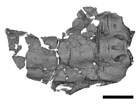
Research led by American Museum of Natural History scientists has provided a fresh perspective on a shark that might have been the “Jaws” of the Carboniferous.  The Carboniferous shark, Carcharopsis wortheni was first scientifically described in the mid-19th century and was previously known only from its characteristic serrated teeth and fragments of jaw.  A fossilised brain case identified as C. wortheni was excavated in 2007 from Upper Mississippian aged rocks in Arkansas (Fayetteville Shale).  The fossil was found by Royal Mapes, a retired Ohio University professor and a research associate at the Museum.  State-of-the-art imaging techniques were used to provide a unique insight into this important apex predator that lived some 320 million years ago.

The Fossil Cranium (Brain Case) of Carcharopsis wortheni

The fossilised brain case of C. wortheni.
The brain case of Carcharopsis wortheni. A Carboniferous shark study.

Picture credit: American Museum of Natural History/Allison Bronson

Insight into Shark Evolution

The shark lived at a critical point in the evolutionary history of our planet, part of a marine fauna that survived the end Devonian mass extinction event which decimated vertebrate species.  This ancient Palaeozoic shark,  was originally described in 1843 based on its distinctive serrated teeth, a feature that is common in extant sharks such as the formidable Great White (Carcharodon carcharias), an apex marine predator with a frightening reputation, thanks in the main to the film “Jaws” directed by Steven Spielberg, which was based on Peter Benchley’s book.  However, serrated teeth are rarely found in Palaeozoic sharks.

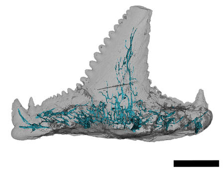
A CT Scan Showing the Unique Serrations on a Tooth from Carcharopsis (C. wortheni)

Shark fossil tooth C. wortheni.
Carcharopsis fossil tooth.  Scale bar = 1 mm.

Picture credit: American Museum of Natural History/Allison Bronson

The picture above shows a computer tomography generated image of a Carcharopsis tooth, measuring around five millimetres in length.  The blue lines are canals identified within the tooth.

Commenting on the fossil, lead author of the study Allison Bronson, a PhD student at the American Museum of Natural History stated:

“They [the teeth] look a little like what you’d see in a Great White, but are 320 million years old and with different enamel.  This is really early to see serrated teeth.”

Carboniferous Shark Fossil Study

Royal Mapes donated the brain case specimen to the New York-based natural history museum, along with a remarkable 540,000 other fossils.  Mapes co-authored the Carcharopsis study, which has been published in the journal “Papers in Palaeontology”, American Museum of Natural History curator John Maisey also contributed to the paper.

The scientists used high-resolution computed tomography (CT) imaging to examine the cranium, a tooth, and an isolated portion of a tooth base.  Using the scans, they were able to reconstruct the internal canals of the teeth for the first time and found that these are similar to the canals found in today’s sharks.

The arrangement of the shark’s blood vessels—also revealed through CT scans—suggests that Carcharopsis was probably closely related to the group of ancient cartilaginous fish from which today’s sharks and rays evolved.  However, more complete fossils are needed to firmly position it in the tree of life.

Everything Dinosaur acknowledges the help of the American Museum of Natural History in the compilation of this article.

Visit the Everything Dinosaur website: Everything Dinosaur.

Go to Top