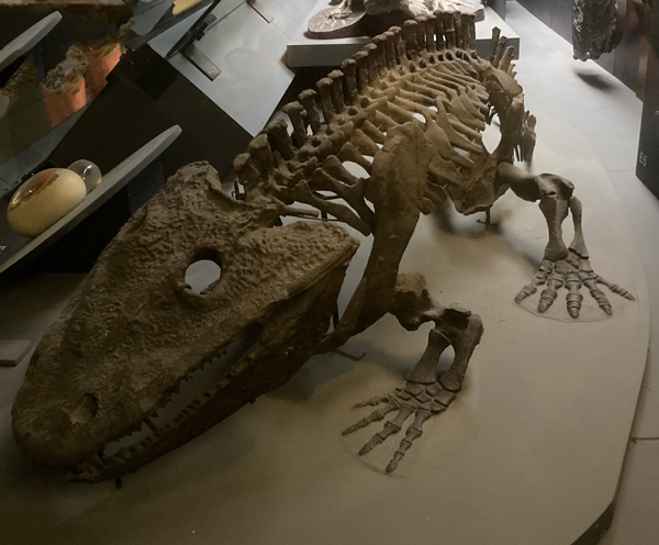
April Fool’s Day is the perfect excuse for a little mischievous fun. Every year on the 1st of April, people play practical jokes and the media is full of bizarre news stories that turn out to be hoaxes. However, while jokes are often harmless fun, some deceptions have had a lasting impact on science. Prehistoric pranks can come in various shapes and sizes. For example, we are aware of bogus new dinosaur model announcements in the past. Strange chimera consisting of theropods crossed with ceratopsians. Indeed, we have been asked to help out with museums when they wanted to add a little bit of mischief to their April communications.
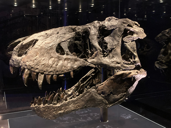
In addition, even the likes of Sir David Attenborough have been fooled by fossils. The accomplished naturalist and broadcaster recalls the story of how he was once convinced to purchase a trilobite mating fossil. The specimen proved to be a fake, a clever concoction prepared by a Moroccan fossil preparator to bump up the price.
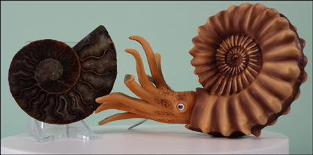
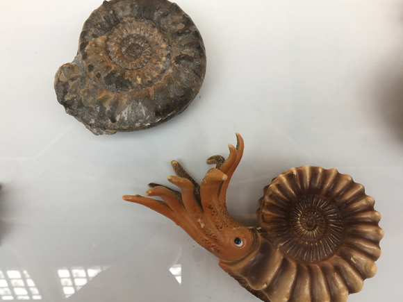
Calymene trilobites. Unscrupulous fossil sellers often “customise” fossils to make them more valuable. Even Sir David Attenborough has been caught out by such practices. Picture credit: Everything Dinosaur.
Picture credit: Everything Dinosaur
When Science Gets it Wrong
In fact, the world of palaeontology has not been immune to prehistoric pranks and trickery. Over the years, a few famous fossil hoaxes have fooled experts and captured the public imagination. So, as we enjoy April Fool’s Day, it is worth exploring how even scientists can sometimes be misled. Science relies on evidence. However, mistakes can be made when it comes to interpreting the evidence. Occasionally, these mistakes are not accidents at all. Instead, they are deliberate hoaxes designed to mislead.
One of the most famous examples is the so-called “missing link” known as Piltdown Man. In 1912, fragments of a skull were discovered in Sussex, England. At the time, this discovery seemed extraordinary. The remains were claimed to represent a new species of early human, one with both human and ape-like features. It was not until 1953 that the fossils were definitively proved to be a hoax. The material was identified as a human skull from the Middle Ages, fossil teeth from ape (chimpanzee), and an orangutan jawbone that was a few hundred years old.
As a result, Piltdown Man became one of the greatest scientific hoaxes of all time.
Lessons from a Prehistoric Prank
The Piltdown Man forgery provides important lessons for palaeontologists. Science is not just about discovery; it is also about verification. Because of this case, scientists became more cautious. They now rely on improved techniques and stricter peer review.
Similarly, a fossil from China claimed to be a missing link between birds and theropod dinosaurs. The specimen was given the informal name of “Archaeoraptor”. A sensational article announced this remarkable discovery in an article in “National Geographic” magazine (1999). Subsequently, the specimen was found to consist of several pieces from real fossils that had been rearranged and stuck together to make the material more valuable.
Moreover, the story highlights how expectations can influence interpretation. At the time, many researchers expected a transitional form between terrestrial theropods and true birds to be discovered.
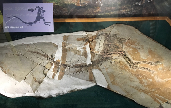
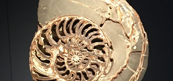
Sinosauropteryx on display – the first feathered dinosaur described. It was described in 1996, and scientists were expecting more feathered dinosaur fossils from China. This helped set the scene for the “Archaeoraptor” forgery. Picture credit: Everything Dinosaur.
Picture credit: Everything Dinosaur
A Light-Hearted Look at Fossil Discoveries
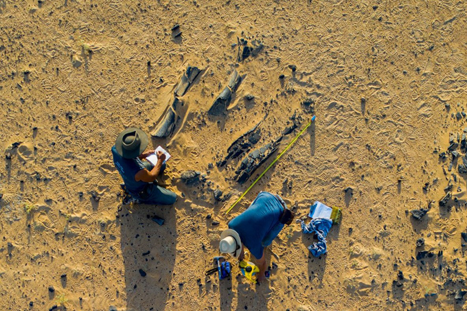
Of course, not all “fossil surprises” are serious. Every year, museums, universities, and even palaeontology blogs join in April Fool’s Day fun. From imaginary dinosaur species to bizarre fossil discoveries, these playful stories entertain and educate at the same time. At Everything Dinosaur, we have enjoyed joining in over the years. After all, a good-natured joke can spark interest in the Earth sciences. For example, fifteen years ago we were asked to help promote a prehistoric animal exhibition by helping to organise the discovery of a dinosaur bone on a beach in Cumbria.
To read more about this prehistoric prank: Unbelievable! Dinosaur Bone Found at Whitehaven.
April Fool’s Day is an annual reminder to question what we see and read. Whether it is a surprising fossil claim or an unusual news story, it is best to be sceptical. At the same time, curiosity remains at the heart of scientific endeavour. By asking questions and testing ideas, we continue to uncover more amazing evidence about life on Earth.
So, if you come across a “new dinosaur discovery” today, take a closer look. It might just be an April Fool’s trick!
And if it is real—well, that’s even more exciting!
The award-winning Everything Dinosaur website: Dinosaur and Prehistoric Animal Models.