Articles, features and information which have slightly more scientific content with an emphasis on palaeontology, such as updates on academic papers, published papers etc.
29
07, 2026
Newly Described Sauropodomorph from the Late Triassic of Zimbabwe
A team of international researchers have described a new dinosaur based on fossils found in Zimbabwe. This newly described sauropodomorph named Musango matusadonaensis lived during the Late Triassic (Norian faunal stage), around 210 million years ago. Musango is the fifth dinosaur to be named from fossils discovered in Zimbabwe. Furthermore, these fossils further support the idea of distinct dinosaur communities in southern Africa during the Late Triassic. Previously, scientists had thought that there were close faunal and biostratigraphical links between the Late Triassic vertebrate faunas of Zimbabwe and South Africa. Currently, no dinosaur taxa are shared between the Triassic-aged sedimentary units in Zimbabwe and those of the main Karoo Basin of South Africa and Lesotho.

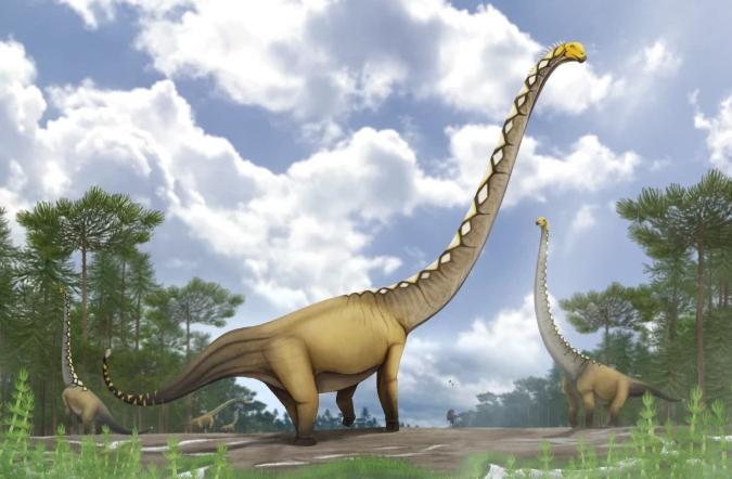
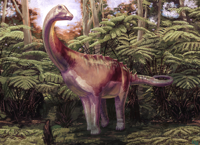
A life reconstruction of the fifth dinosaur to be named from fossils found in Zimbabwe (Musango matusadonaensis). Picture credit: Mark Witton.
Picture credit: Mark Witton
The fossils were discovered on the shores of Lake Kariba in northern Zimbabwe. Moreover, the find provides fresh evidence that southern Africa preserves an important record of early dinosaur evolution.
Musango matusadonaensis – A New Member of the Sauropodomorpha
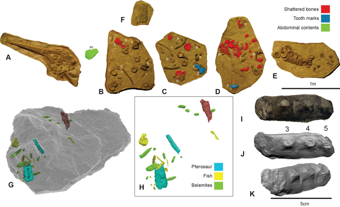
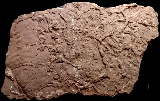
Musango matusadonaensis is classified as an early member of the Sauropodomorpha, a group of dinosaurs that eventually gave rise to giant, long-necked herbivores such as Brachiosaurus and Diplodocus. However, unlike its enormous descendants, M. matusadonaensis was a relatively small, lightly built dinosaur that probably walked on two legs. The fossils were found in association with each other but not articulated. The material consists of a partial skeleton consisting of five dorsal vertebrae, two sacral vertebrae, a left scapula and coracoid along with a partial left ilium; a right pubis and elements from the limbs. In addition, several indeterminate bone fragments were recovered.
The absence of a femur makes size estimation difficult. However, based on comparisons with other Late Triassic sauropodomorphs, the specimen (NHMZ 2583) represents a four and a half metre long individual. This dinosaur is estimated to have weighed around 220 kilograms.

Musango matusadonaensis silhouette showing known fossil material. Scale bar equals 1 metre. Picture credit: Brandon Stuart with additional annotation by Everything Dinosaur.
Picture credit: Brandon Stuart with additional annotation by Everything Dinosaur
While the skull has not yet been found, researchers believe that Musango was probably herbivorous or perhaps omnivorous.
Revealing Ancient African Ecosystems
The Late Triassic was a pivotal period in Earth’s history. Dinosaurs had evolved, but they had not yet become the dominant terrestrial vertebrates they would be during the Jurassic and Cretaceous. For many years, scientists assumed that dinosaur faunas across southern Africa were broadly similar. However, discoveries from Zimbabwe are beginning to challenge this long-held view.
Professor Paul Barrett, Merit Researcher at the Natural History Museum, London, and the study’s lead author, commented:
“Until recently, it was assumed that the dinosaurs living across southern Africa were largely the same. However, these discoveries are showing that this part of the ancient supercontinent Gondwana was actually made of a series of smaller ecosystems – each with a different cast of characters.”

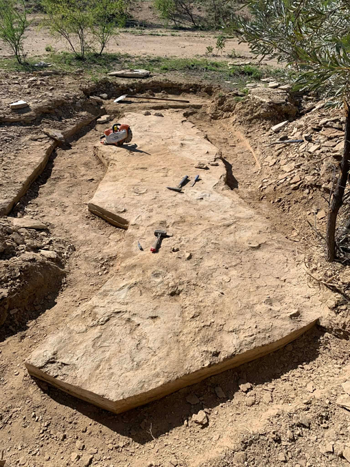
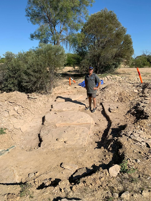
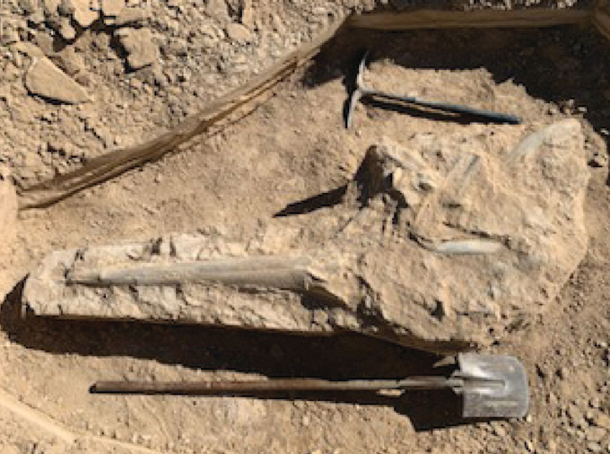
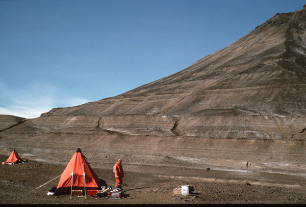
Professor Paul Barrett (London Natural History Museum) carefully excavating fossil material on the shores of Lake Kariba. Picture credit: Paul Barrett.
Picture credit: Paul Barrett
The Fifth Dinosaur to be Described from Zimbabwe
The first dinosaur to be named from Zimbabwe was the coelophysoid “Syntarsus” rhodesiensis which was formally named and described in 1969. However, the genus name was already assigned, so it was moved to the genus Megapnosaurus, although the fossils may represent a species of Coelophysis.
The five dinosaurs known from Zimbabwe:
- “Syntarsus” – Megapnosaurus (possibly Coelophysis) – a small theropod dinosaur.
- Vulcanodon karibaensis – a primitive sauropod.
- Mbiresaurus raathi – a basal sauropodomorph named in 2022 that is geologically older than Musango matusadonaensis (Griffen et al).
- Musankwa sanyatiensis – a basal sauropodomorph named in 2024 (Barrett et al) which was coeval with M. matusadonaensis.
- Musango matusadonaensis (Barret et al, 2026).
To read our article from 2024 about Musankwa sanyatiensis, the fourth dinosaur to be described from Zimbabwe: A New Sauropodomorph from Zimbabwe (2024).
The discovery of several sauropodomorphs in this locality highlights the significance of southern Africa to researchers examining the evolution of the sauropod lineage.
Analysis of the Musango fossil bones indicates that the individual was around eight years old and approaching full adult size when it died. Interestingly, the skeleton also preserves evidence that the dinosaur survived a serious injury or infection during its lifetime. This suggests it recovered successfully before eventually dying from another cause.
Life in an Ancient River Ecosystem
During the Late Triassic, the landscape of present-day Zimbabwe looked very different from today. Rivers and streams crossed the region, supporting a wide range of plants and animals. Musango matusadonaensis shared its habitat with lungfish, crocodile-like phytosaurs and other dinosaurs, including Musankwa sanyatiensis. As fieldwork continues, researchers expect many more species to emerge from these fossil-rich rocks.
Professor Paul Barrett believes these discoveries are only the beginning:
“It suggests that what we’ve found so far is only the tip of the iceberg. We’ve already got one other new species that we’ve yet to describe, while our team has been told about other fossils from the region that we’ve yet to look at. I think it’s likely there are even more dinosaurs still to be found there.”

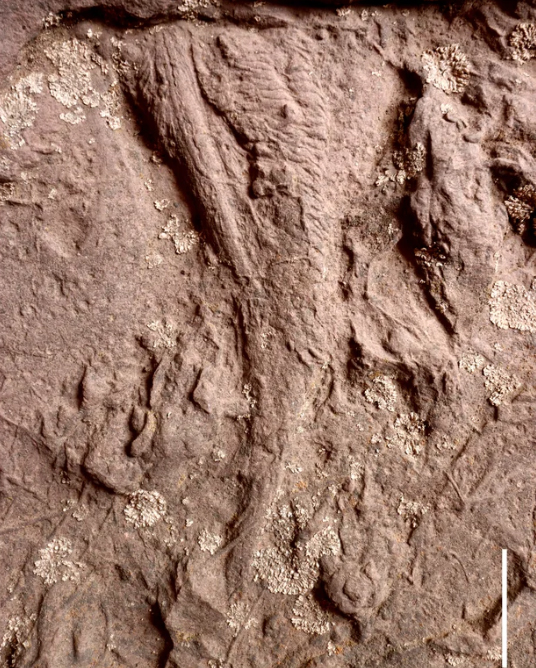
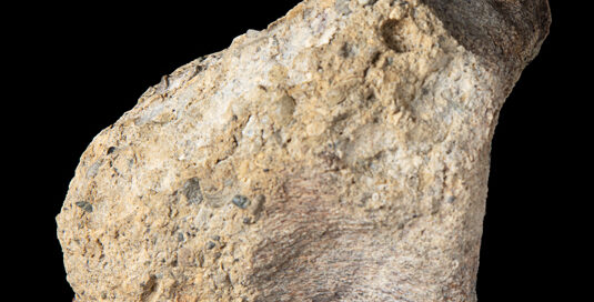
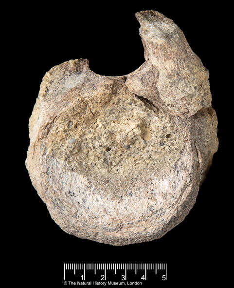
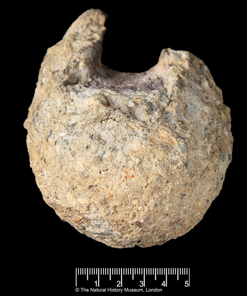
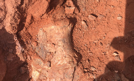
The ilium of the newly described sauropodomorph from Zimbabwe (Musango matusadonaensis). Picture credit: Brandon Stuart.
Picture credit: Brandon Stuart
An International Scientific Collaboration
The research forms part of an ongoing collaboration between scientists from Zimbabwe, South Africa and the United Kingdom. Together, they are exploring regions that have received relatively little palaeontological attention compared to Europe, China and North America. As a result, these discoveries are helping fill important gaps in the fossil record. They also demonstrate that Africa played a significant role in the early evolution and diversification of dinosaurs.
Professor Jonah Choiniere, leader of the expedition and a co-author from Johannesburg’s Evolutionary Studies Institute, explained:
“New dinosaur species like Musango show the value of doing palaeontological fieldwork in remote, and often scenically beautiful, places. This study is the result of a thriving international collaboration between the UK, South Africa and Zimbabwe, and reinforces the importance of southern Africa in understanding dinosaur diversity.”
Musango matusadonaensis Etymology
The name of this new dinosaur reflects both the local language and the discovery site. Musango comes from the ChiShona language and means “living in the bush”, referring to the remote location where the dinosaur was unearthed. Meanwhile, the species name honours the nearby Matusadona National Park.
The discovery of the fifth dinosaur from Zimbabwe adds another important piece to the puzzle of dinosaur evolution. Moreover, it highlights the scientific importance of the country’s fossil record. As exploration continues, palaeontologists hope that many more prehistoric species will emerge from this remarkable part of Gondwana.
Everything Dinosaur acknowledges the assistance of a media release from the London Natural History Museum in the compilation of this article.
The scientific paper: “A new sauropodomorph dinosaur from the Pebbly Arkose Formation (Upper Triassic: Norian) of Kariba, Zimbabwe” by Paul M. Barrett, Jennifer Botha, Lara Sciscioe, Brandon P. Stuart, Jack Lovegrove, Darlington Munyikwa, Michel Zondo, Timothy J. Broderick, Steve F. Edwards, Edward Mbambo, Kimberley E. J. Chapelle, Kathleen N. Dollman, Steve Tolan and Jonah N. Choiniere published in the Journal of Systematic Palaeontology.
The award-winning Everything Dinosaur website: Sauropod Models and Dinosaur Figures.