Fossil finds, new dinosaur discoveries, news and views from the world of palaeontology and other Earth sciences.
3
05, 2022
Getting your Claws into Therizinosaurs
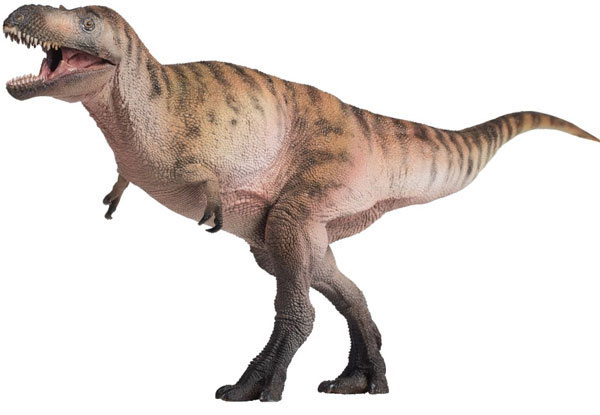
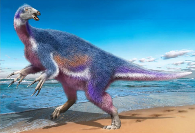
Scientists have named a new species of therizinosaur based on fragmentary remains found on the Japanese island of Hokkaido. The dinosaur has been named Paralitherizinosaurus japonicus, it is the first recovered from Asian marine deposits and the third example of a therizinosaur to be found in Japan.
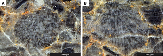
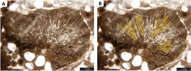
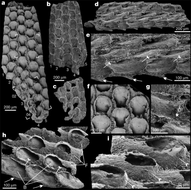
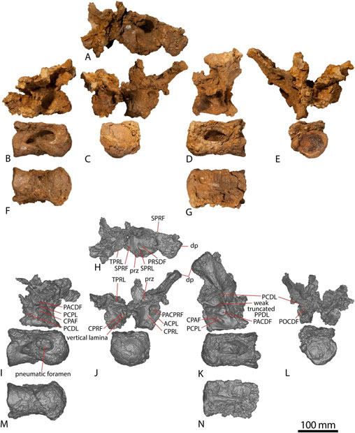
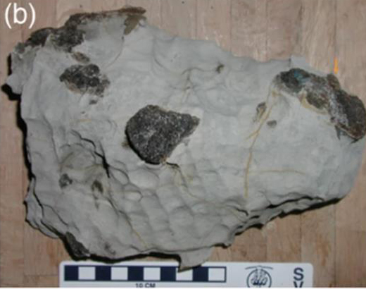
The fossil material, recovered from the lower Campanian Osoushinai Formation near to the town of Nakagawa in the Hokkaido Prefecture, was previously identified as a maniraptoran theropod dinosaur, possibly therizinosaur, but its taxonomic status remained uncertain. A group of scientists including Yoshitsugu Kobayashi and Anthony R. Fiorillo from the Hokkaido University Museum re-examined the fossils and erected a new taxon confirming the fossil material did represent a Late Cretaceous member of the Therizinosauridae.
Evolution of Claw Shape in the Therizinosauridae
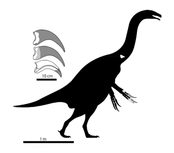
Writing in the academic journal “Scientific Reports”, the researchers reassessed the fossil material consisting of a single vertebra plus bones and claws (unguals) from the right hand. As well as concluding that the fossils represent a therizinosaur, they confirmed that it is the geologically youngest therizinosaur known from Japan described to date.
Important Implications for Claw (Ungual) Evolution in the Therizinosauridae
The scientists compared the shape of the hand claws from Paralitherizinosaurus japonicus with the claws from geologically older therizinosaurs and they postulated that that primitive therizinosaurs had claws with generalist functionalities and that the claws of more derived, later therizinosaurs such as P. japonicus were more suited to the hook-and-pull feeding function. Hook-and-pull feeding involves the use of the claws to help gather vegetation and bring it closer to the mouth.
What’s in a Name?
The fossils were found in a concretion associated with the Campanian-aged Osoushinai Formation of the Yezo Group on Hokkaido Island. The Yezo Group mostly consists of marine deposits and many vertebrate fossils such as plesiosaurs, sharks, mosasaurs and turtles have been discovered. Fragmentary dinosaur fossils are also associated with these strata including hadrosaurids, an armoured dinosaur (nodosaurid) and a potential tyrannosaur. A therizinosaur taxon can now be added to this Late Cretaceous dinosaur biota.

To view replicas of feathered theropods including therizinosaurus (whilst stocks last): PNSO Age of Dinosaurs Figures.
The discovery of the bones and claw elements in marine deposits helped to inspire this dinosaur’s scientific name. The genus name translates as “scythe reptile by the sea”, whilst the species name honours Japan.
The scientific paper: “New therizinosaurid dinosaur from the marine Osoushinai Formation (Upper Cretaceous, Japan) provides insight for function and evolution of therizinosaur claws” by Yoshitsugu Kobayashi, Ryuji Takasaki, Anthony R. Fiorillo, Tsogtbaatar Chinzorig and Yoshinori Hikida published in Scientific Reports.