Pictures of fossils, fossil hunting trips, fossil sites and photographs relating to fossil hunting and fossil finds.
29
12, 2022
A New Dwarf Nodosaurid Called Patagopelta
In today’s blog post we look at the dwarf nodosaurid Patagopelta (P. cristata), which was formally named and described earlier this month.
A new, very small, armoured dinosaur has been named and described from fossils found in Argentina. The dinosaur which measured around 2 to 2.3 metres in length (based on the dimensions of the femur), suggests that some members of the Nodosauridae in Gondwana became smaller in the Late Cretaceous, perhaps as armoured dinosaurs in South America were under evolutionary pressure from other ornithischians and titanosaurs.

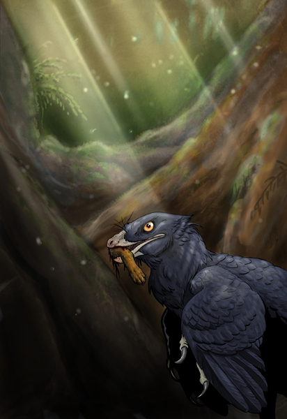
Dwarf Nodosaurid Patagopelta
Fragmentary remains of Late Cretaceous armoured dinosaurs are known from Chile and Argentina, but little work had been undertaken to assess these specimens and to review their phylogeny and taxonomic relationship with other members of the Ankylosauria clade from North America and elsewhere in the world.
Writing in the ” Journal of Systematic Palaeontology”, the researchers led by Facundo Riguetti, a CONICET doctoral fellow, reassessed the known ankylosaur material in conjunction with some other recently found fossils and, as a result, they were able to establish a new nodosaurid species from bones and a single tooth found in sediments of the Allen Formation (Campanian–Maastrichtian) in Salitral Moreno, Río Negro Province (northern Patagonia).
Patagopelta cristata
The dinosaur’s genus name translates as “Patagonian shield” whilst the trivial name derives from the Latin for crest – a reference to the diagnostic crests on both the anterior surface of the femur and the lateral osteoderms of the cervical rings.
Dr Riguetti commented:
“The importance of the study lies in the fact that Patagopelta is the first species of Ankylosauria described for the continental territory of Argentina, which fills the existing gap for this group and adds a new thyreophoran to the very few incomplete and indeterminate remains known for our country from this type of ornithischian dinosaur.”
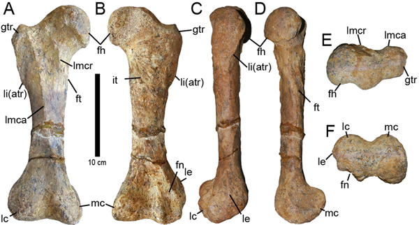
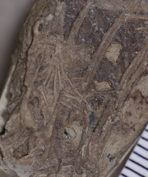
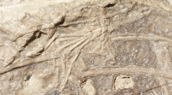
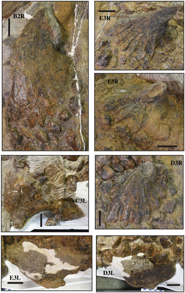
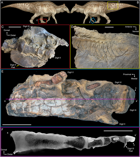
The Right Femur
The best-preserved fossil element is the right femur, which is complete and shows typical anatomical characteristics associated with the Nodosauridae. This bone along with the distinctive cervical osteoderms led to the erection of this new species. As the femur is only 25 cm in length and bone histology suggests an adult animal, the researchers conclude that Patagopelta was a dwarf form of armoured dinosaur.
Co-author Sebastián Apesteguía, a CONICET researcher, explained:
“For an armoured dinosaur, Patagopelta is extremely small. Due to the size of the femur, only 25 centimetres in length, we estimate that the animal must have been between two and three meters long, while, in general, ankylosaurs are medium-sized or large animals, with an average length of between four and five metres.”
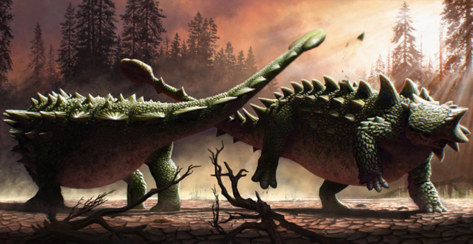
A Faunal Exchange Across the Americas
Although it is thought that the Nodosauridae evolved in the Northern Hemisphere, towards the end of the Cretaceous (Campanian – Maastrichtian), a land bridge existed between North America and South America that permitted a faunal exchange. Titanosaurs migrated north, which explains why fossils of titanosaurs such as Alamosaurus occur in the USA. Ornithischian dinosaurs such as hadrosaurs and nodosaurids moved south.
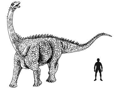
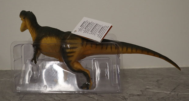
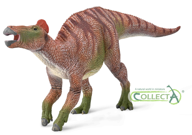
The image above shows a typical Late Cretaceous titanosaur, for models of Late Cretaceous dinosaurs including titanosaurs and armoured dinosaurs: CollectA Prehistoric Life Models.
Sebastián Apesteguía added:
“That is why in South America we only expect to find animals like Patagopelta in rocks from the Late Cretaceous, just before the global extinction of the dinosaurs took place.”
Dwarfism in Late Cretaceous South American Thyreophora
The size of Patagopelta along with the recently described Stegouros (Soto-Acuña et al, 2021)*, from southernmost Chile, suggests that armoured dinosaurs in South America may have gradually become smaller. This trait is not known in members of the Thyreophora described from other parts of the world. Palaeontologists have speculated that perhaps competition from titanosaurs and the migration of hadrosaurs into South America might have led to armoured dinosaurs adapting to different ecological niches to avoid competition. By being smaller these animals needed fewer resources than larger, contemporaneous herbivorous dinosaurs.
It has also been suggested that the geology of Patagonia where the fossils of Patagopelta were found might provide a clue to the dwarfism. Geologists are aware of several Late Cretaceous marine transgressions in the region. This might have led to the establishment of an island archipelago with dinosaurs living on these small islands gradually become smaller due to a scarcity of resources (the “island rule”).
Tracks of Dwarf Ankylosaurs
Members of the Patagopelta research team had previously described tracks of dwarf ankylosaurs, possibly affected by similar circumstances, preserved in Upper Cretaceous deposits in Bolivia.
*To read Everything Dinosaur’s 2021 article about the discovery of Stegouros: New Armoured Dinosaur from Chile.
Everything Dinosaur acknowledges the assistance of a media release from Consejo Nacional de Investigaciones Científicas y Técnicas (CONICET) in the compilation of this article.
The scientific paper: “A new small-bodied ankylosaurian dinosaur from the Upper Cretaceous of North Patagonia (Río Negro Province, Argentina)” by Facundo Riguetti, Xabier Pereda-Suberbiola, Denis Ponce, Leonardo Salgado, Sebastián Apesteguía, Sebastián Rozadilla and Victoria Arbour published in the Journal of Systematic Palaeontology.