All about dinosaurs, fossils and prehistoric animals by Everything Dinosaur team members.

Articles, features and information which have slightly more scientific content with an emphasis on palaeontology, such as updates on academic papers, published papers etc.

14 12, 2022

Tail Clubs for Social Dominance

By |2023-02-07T09:29:08+00:00December 14th, 2022|Categories: Dinosaur and Prehistoric Animal News Stories, Dinosaur Fans, Main Page, Palaeontological articles, Photos/Pictures of Fossils|0 Comments

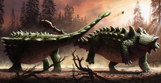
Ankylosaurs battled each other using their tail clubs for social dominance in intraspecific combat. A recently published scientific paper on the ankylosaur Zuul crurivastator suggests that these armoured dinosaurs used their tail clubs to bash each other as well as to fend off tyrannosaurs.

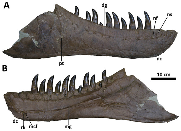
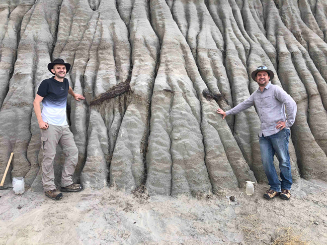
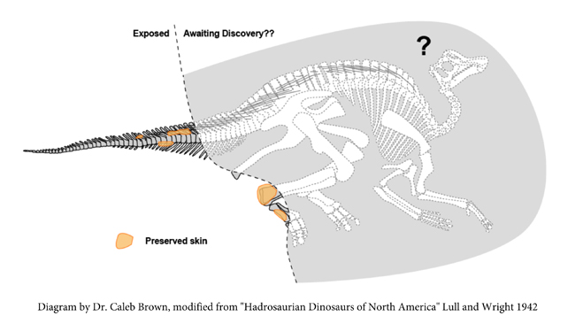
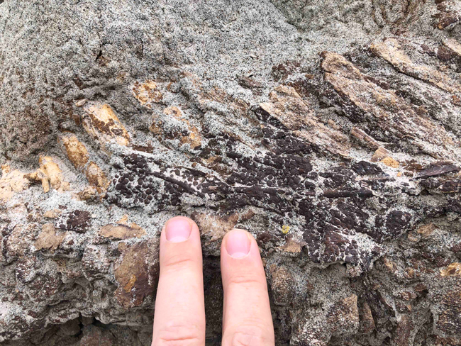
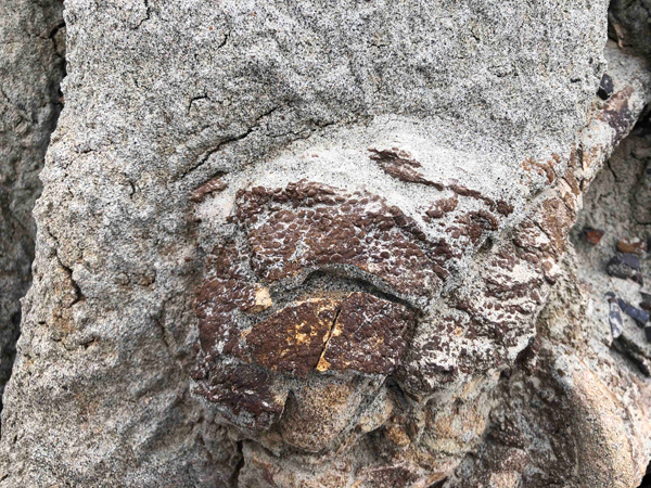
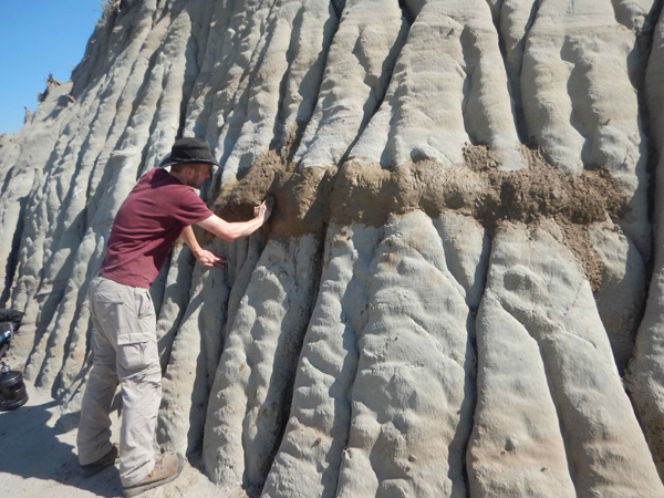
In the study, published in “Biology Letters” the research team, examined the osteoderms of the remarkably well preserved Zuul crurivastator, an armoured dinosaur described from fossils found in the Coal Ridge Member of the Judith River Formation (Montana). Several of osteoderms along the flanks show signs of damage and healing which led the scientists to postulate that these dinosaurs battled each other with their tail clubs. These fights would have been for social or territorial dominance, perhaps even a result of a “rutting” season fighting for mates – behaviour associated with many mammals today.

Tail clubs for social dominance.
A pair of ankylosaurs (Zuul crurivastator) indulge in some intraspecific combat. Picture credit: Henry Sharpe

Zuul crurivastator

Named and formally described in 2017, Zuul crurivastator (pronounced Zoo-ul cruh-uh-vass-tate-or) roamed the northern part of Laramidia approximately 76 million years ago (Campanian faunal stage of the Late Cretaceous).

To read Everything Dinosaur’s 2017 blog post about the fossil discovery: Zuul – The Destroyer of Shins.

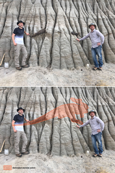
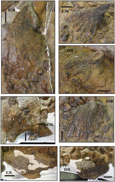
Zuul’s body was covered in bony plates (osteoderms) of different shapes and sizes and the ones along its flanks were particularly large and spiky. Interestingly, the scientists which included lead author and renowned ankylosaur expert Dr Victoria Arbour (Royal British Columbia Museum, Canada), noted that dermal armour near the hips on both sides of the body showed damage that had subsequently healed. This localised, bilaterally symmetrical pathology is speculated to have been caused by ritualised combat rather than wounds inflicted by an attacking theropod dinosaur.

Damaged osteoderms on the Zuul Holotype
Identifying damaged osteoderms in the holotype of Zuul crurivastator. A composite photograph of the skull, first cervical half ring, body block and tail block (top). Fossil material is brown and surrounding rock matrix is grey. Interpretive illustration showing non-pathological osteoderms in white and pathological osteoderms in red (bottom). Picture credit: Arbour, Zanno and Evans.

An Exciting Piece of the Ankylosaur Puzzle

Dr Arbour commented:

“I’ve been interested in how ankylosaurs used their tail clubs for years and this is a really exciting new
piece of the puzzle. We know that ankylosaurs could use their tail clubs to deliver very strong blows to an opponent, but most people thought they were using their tail clubs to fight predators. Instead, ankylosaurs like Zuul may have been fighting each other.”

The genus name honours a fictional monster from the 1984 film “Ghostbusters”, whilst the trivial part of the binomial name translates as “the destroyer of shins”, a nod to the idea that tail clubs were used as defensive weapons to deter attacks from predatory theropod dinosaurs. The substantial club on the end of the three-metre-long tail being used to bash into the lower legs of tyrannosaurs. This new research does not refute the idea that these tail clubs had a role in defence, but based on the pathology seen in the Zuul holotype (specimen number ROM 75860) the scientists propose that sexual selection and intraspecific combat drove their evolution. Many mammals today such as deer, antelope, cattle and sheep have horns and antlers that have evolved for use in battles between members of the same species.

Damaged osteoderms in an ankylosaur.
Details of pathological and non-pathological osteoderm morphology in ROM 75860 (Zuul crurivastator). B2R and E3R are non-pathological flank osteoderms. F3R and D3R are pathological flank osteoderms missing the tips of the apex, and the keratinous sheath has not grown over the tip. D3L is a pathological flank osteoderm missing a large portion of the apex, and the keratinous sheath has overgrown the damaged region. C3L and E3L are pathological flank osteoderms with highly modified morphologies, missing large portions of the trailing posterior edge and with the keratinous sheath covering the damaged region. Picture credit: Arbour, Zanno and Evans.

It had been suggested previously that ankylosaurs may have clubbed each other, and that broken and healed ribs could provide evidence to support this idea. Unfortunately, ankylosaurid skeletons are extremely rare in the fossil record, these animals were not common, even in the Late Cretaceous of North America, where the ecosystem was dominated by other ornithischian dinosaurs such as duck-billed dinosaurs and ceratopsians.

Implications for Ankylosaur Behaviour

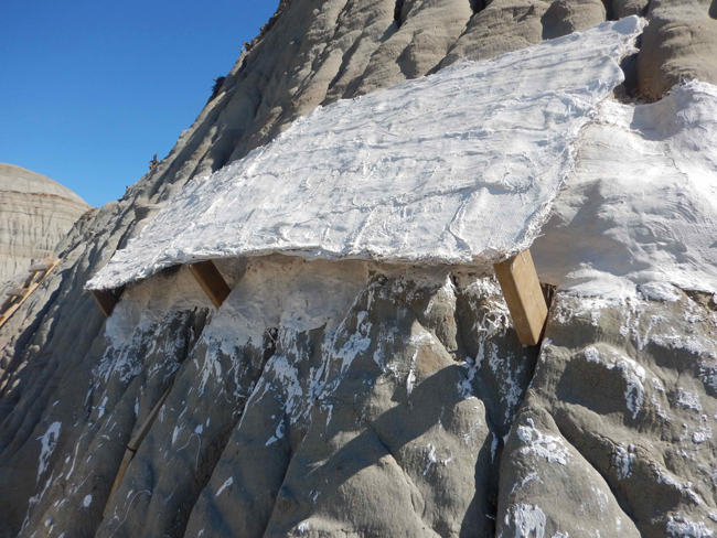
The remarkable Zuul fossil skeleton provides palaeontologists with an opportunity to study pathology recorded on the bones and dermal armour.

Co-author Dr David Evans (Curator of Vertebrate Palaeontology at the Royal Ontario Museum) explained:

“The fact that the skin and armour are preserved in place is like a snapshot of how Zuul looked when it
was alive. And the injuries Zuul sustained during its lifetime tell us about how it may have behaved and
interacted with other animals in its ancient environment.”

Tail Clubs for Social Dominance

The researchers conclude that the imposing tail club of Zuul could have been used in defence when needed, but the analysis suggest that sexual selection drove the evolution of this weapon. This finding has consequences for how palaeontologists perceive ankylosaurs. It suggests that these dinosaurs were capable of complex behaviours and that they likely engaged in ritualised combat over mates or for social dominance as inferred in other types of dinosaurs and observed in living mammals and birds.

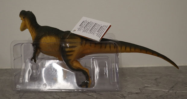
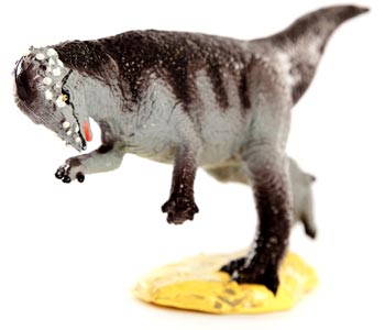
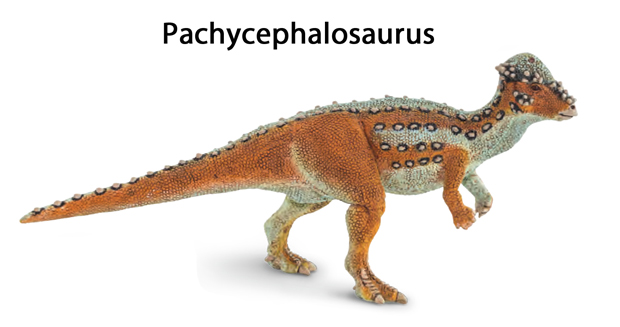
To view a replica of the armoured dinosaur Zuul and other prehistoric animal models (whilst stocks last): Armoured Dinosaurs and Prehistoric Animal Figures (Wild Safari).

Everything Dinosaur acknowledges the assistance of a media release from the Royal Ontario Museum in the compilation of this article.

The scientific paper: “Palaeopathological evidence for intraspecific combat in ankylosaurid dinosaurs” by Victoria M. Arbour, Lindsay E. Zanno and David C. Evans published in Biology Letters.

13 12, 2022

Spinosaurus Not an Aquatic Dinosaur According to New Research

By |2024-02-25T07:41:24+00:00December 13th, 2022|Categories: Adobe CS5, Dinosaur and Prehistoric Animal News Stories, Dinosaur Fans, Main Page, Palaeontological articles|1 Comment

A recently published scientific paper has refuted the idea that Spinosaurus (S. aegyptiacus) was an aquatic dinosaur. Writing in the academic journal eLife, the researchers which included Paul Sereno (University of Chicago), conclude that Spinosaurus was not aquatic. Instead, they revert to the earlier hypothesis that this super-sized carnivore was a semi-aquatic, bipedal predator that did feed on fish but ranged far inland.

Over a decade ago, Everything Dinosaur team members were contacted by members of the CGI team working on the Spinosaurus segment of the soon to be launched television series entitled “Planet Dinosaur”. At the time, (2010), S. aegyptiacus was thought to be a semi-aquatic, bipedal predator that specialised in hunting fish, but was not thought to be an almost entirely aquatic dinosaur.

The front cover of the book "Planet Dinosaur"
From paddler to swimming and back to paddler again – the “evolving” image of Spinosaurus. A recently published paper refutes the hypothesis that Spinosaurus aegyptiacus was an aquatic animal. Researchers postulate that it was semi-aquatic, a biped feeding on fish but not a dinosaur that could dive and swim very efficiently. Picture credit: BBC Worldwide.

Picture credit: BBC Worldwide

The 2014 Scientific Paper

In 2014, Paul Sereno (the lead author of the latest paper), was one of the authors of a study into Spinosaurus material found in Morocco. In the paper, the researchers proposed that this huge theropod possessed adaptations that indicated a semi-aquatic lifestyle. The skull had small nostrils located further up the snout to allow this dinosaur to breathe whilst its jaws were partially submerged and neurovascular openings along the jaws were interpreted to be sense organs that permitted Spinosaurus to sense the movements of fish through the water. Analysis of the centre of gravity indicated that Spinosaurus was a quadruped.

To read Everything Dinosaur’s blog post about the 2014 study: Spinosaurus – Four Legs are Better than Two!

However, commenting on the 2014 paper, Professor Sereno admits that there was a mistake made when examining the centre of gravity of Spinosaurus. When the centre of gravity was calculated leading to the conclusion that this huge theropod walked on all fours, the volume of the lungs were not properly accounted for. When a new assessment of the centre of gravity is made, using a more accurate lung volume assessment, the results are radically different. The data suggests that Spinosaurus was a biped, with a typical posture associated with other large-bodied carnivorous dinosaurs.

Spinosaurus model.
The new research which included a more accurate assessment of lung volume, indicates that Spinosaurus was bipedal, perhaps the Jurassic Park III Spinosaurus was not that inaccurate after all?

The image (above) depicts Spinosaurus with the typical bipedal posture of a large-bodied theropod dinosaur. The model is a Nanmu Studio Spinosaurus (version 2.0).

To view the range of Nanmu Studio models and figures available from Everything Dinosaur: Nanmu Studio Dinosaur and Prehistoric Animal Replicas.

The Fleshy Spinosaurus Tail

In 2020, another paper was published which examined the caudal vertebrae of Spinosaurus aegyptiacus, tail bones having not been part of previous studies. Writing in the journal “Nature” the researchers which included Nizar Ibrahim (University of Detroit Mercy, Detroit), along with colleagues from the University of Portsmouth, proposed that Spinosaurus had a broad, flexible, fin-like tail that could have propelled this dinosaur through the water. This seemed to be the decisive evidence, that Spinosaurus was indeed an aquatic hunter.

To read Everything Dinosaur’s blog post about the 2020 paper: Spinosaurus – The River Monster.

Spinosaurus was not Aquatic

In this newly published research, Sereno and his colleagues looked at the biomechanics of the fin-like tail and analysed its effectiveness as an organ of propulsion through water. When compared to alligators, the tail and hind feet of Spinosaurus were found to be very inefficient swimming organs. Spinosaurus was an unstable, slow-surface swimmer only capable of a swimming speed of less than one metre a second.

Spinosaurus tail bones compared to semi-aquatic and aquatic vertebrate caudal vertebrae.
Comparing the skeleton of S. aegyptiacus with a basilisk lizard and aquatic vertebrates. The tail of S. aegyptiacus (A) showing the overlap of neural spines (red) with more posterior caudal vertebrae. A CT scan showing the sail structure of a green basilisk lizard (B). Researchers propose that the tail of Spinosaurus was not adapted to propulsion in water but most probably a display structure. Skeletal illustrations of the caudal vertebrae of aquatic vertebrates (C) – crested newt, a mosasaur, crocodilian and a whale. A graph plotting the centrum proportions along the tail (D) comparing S. aegyptiacus with a crested newt (Triturus cristatus), semi-aquatic lizards (marine iguana Amblyrhynchus, common basilisk Basiliscus basiliscus, Australian water dragon Intellagama lesueurii and a sailfin lizard Hydrosaurus amboinensis), a mosasaur and an American alligator (Alligator mississippiensis). Picture credit: Sereno et al.

The team also calculated that Spinosaurus would have been too buoyant to submerge fully. It was not capable of diving and those robust, heavy hind limbs helped it to walk on land or wade, not acting as additional ballast to help this fifteen-metre-long giant remain underwater.

Sereno and his fellow authors suggest that living reptiles with similar tail bone morphology, such as the basilisk lizard, do not use their tails for propulsion, instead they have a display function.

Spinosaurus Ranged Far Inland

Fossils ascribed to Spinosaurus recovered from fluvial deposits in Niger suggest that this dinosaur ranged far inland. It is true that most Spinosaurus fossils come from sediments that represent extensive coastal deltas. However, these deposits include a large number of non-spinosaurid dinosaur remains, all of which may have been transported for some distance downstream. Spinosaurus fossil material may have also been transported, leading to the misconception that this was a dinosaur confined to the coast. Recently discovered fossils ascribed to Spinosaurus from two inland basins in Niger (Égaro North), indicate that Spinosaurus lived far from the shore. These fossils which include part of an upper jaw (maxilla) were found in fluvial deposits in association with rebbachisaurid and titanosaurian sauropods which are regarded as entirely terrestrial animals.

Spinosaurus was not aquatic
Spinosauridae fossil localities mapped (Early Cretaceous Albian faunal stage). The map (A) shows the fossil location for Baryonyx and Suchomimus (baryonychines) along with spinosaurs (Ichthyovenator, Vallibonavenatrix, Oxalaia, Irritator/Angaturama and Spinosaurus). Yellow asterisks indicate location of Spinosaurus fossils, these range from coastal sites (1 – Bahariya, Egypt and 2 – Zrigat, Morocco) to a site much further inland (3 – Égaro North, Niger). Spinosaurus sp. right maxilla (B), specimen number MNBH EGA1 from central Niger, in medial and ventral views. The inset image shows the fossil material superimposed on the snout of S. aegyptiacus. Abbreviations: am, articular rugosities for opposing maxilla; aofe, antorbital fenestra; Ba, Baryonyx walkeri; en, external naris; Ic, Ichthyovenator laosensis; Ir, Irritator challengeri/Angaturama limai; m3, 12, maxillary alveolus 3, 12; Ox, Oxalaia quilombensis; Su, Suchomimus tenerensis; t, tooth; Va, Vallibonavenatrix cani. Scale bar is 10 cm. Picture credit: Sereno et al.

Comfortable in Water but Not Truly at Home in an Aquatic Environment

Confirming that the researchers think Spinosaurus was a bipedal, semi-aquatic animal that specialised in hunting fish, Professor Sereno added:

“Do I think this animal would have waded into water on a regular basis? Absolutely, but I do not think it was a good swimmer or capable of full submergence behaviour.”

Everything Dinosaur acknowledges the assistance of a media release from the University of Chicago in the compilation of this article.

The scientific paper: “Spinosaurus is not an aquatic dinosaur” by Paul C Sereno, Nathan Myhrvold, Donald M Henderson, Frank E Fish, Daniel Vidal, Stephanie L Baumgart, Tyler M Keillor, Kiersten K Formoso and Lauren L Conroy published in eLife.

8 12, 2022

New Daspletosaurus Species Named

By |2022-12-10T18:25:51+00:00December 8th, 2022|Categories: Adobe CS5, Dinosaur and Prehistoric Animal News Stories, Dinosaur Fans, Main Page, Palaeontological articles, Photos/Pictures of Fossils|0 Comments

A new species of North American, Late Cretaceous tyrannosaur has been announced. It is the third species in the Daspletosaurus genus. Daspletosaurus wilsoni is older than D. horneri, but geologically younger than D. torosus. The skull demonstrates a mix of traits found in primitive tyrannosaurs as well as several more derived characteristics associated with later members of the Tyrannosauridae such as Tyrannosaurus rex.

Writing in the on-line, open access journal PeerJ, the researchers from the Badlands Dinosaur Museum (North Dakota), suggest that the discovery of a tyrannosaur intermediate in age between the previously described species of this genus, supports earlier research identifying several species of Daspletosaurus as a single evolving lineage, and supports the descent of T. rex from this group.

Daspletosaurus wilsoni
Changes in the Daspletosaurus genus over time with three distinct species now identified. Known skull bones of D. wilsoni shown in white. The evolutionary changes in these tyrannosaurs with key anatomical differences listed. Note scale bar = 10 cm. Picture credit: Warshaw and Fowler.

From the Judith River Formation (Campanian Faunal Stage)

The fossil specimen was discovered in 2017. It consists of a partial, disarticulated skull and jaw bones with an exceptional degree of preservation along with vertebrae, a rib and the first metatarsal. The material comes from Judith River Formation exposures in north-eastern Montana. The fossil was found by John Wilson and the species name has been erected in his honour.

The new species displays a mix of characteristics found in more primitive, geologically older tyrannosaurs. For example, D. wilsoni sports a prominent set of horns around the eye, as well as features otherwise known from later members of this group (including T. rex), like a tall eye socket and expanded air-pockets in the skull. In this way, this newly described species represents a transitional form between older and younger tyrannosaur species.

Daspletosaurus wilsoni dentary.
The right dentary of BDM 107 (the holotype). Picture credit: Warshaw and Fowler.

Daspletosaurus wilsoni

Several tyrannosaurs have now been named from the Upper Cretaceous of North America. The researchers note that many of the species are very closely related, forming consecutive sequences where one species evolves into another species over time. When one ancestral species evolves into a second descendant species, this is referred to as anagenesis, as opposed to cladogenesis when successive branching events produce many species that are closely related to each other but not direct descendants of a single species.

The authors of the scientific paper conclude that anagenesis might be the driver of evolutionary change within the Dinosauria at the end of the Cretaceous rather than cladogenetic evolutionary models.

Daspletosaurus Replicas

The first species of Daspletosaurus (D. torosus) was named and described in 1970, the second species, the geologically youngest species, D. horneri was named in 2017. Few models of this tyrannosaur existed but in 2013 CollectA added a Daspletosaurus model to their Prehistoric Life model range. More recently (2021), Safari Ltd introduced a Daspletosaurus figure into their Wild Safari Prehistoric World range.

To view the Wild Safari Prehistoric World range of models available from Everything Dinosaur: Safari Ltd Dinosaur and Prehistoric Animal Models.

Wild Safari Prehistoric World Daspletosaurus dinosaur model
The new for 2021 Wild Safari Prehistoric World Daspletosaurus dinosaur model. Picture credit: Everything Dinosaur.

The scientific paper: “A transitional species of Daspletosaurus Russell, 1970 from the Judith River Formation of eastern Montana” by Elias A. Warshaw and Denver W. Fowler published in PeerJ.

6 12, 2022

Modern Lizards in the Triassic

By |2022-12-08T10:20:48+00:00December 6th, 2022|Categories: Dinosaur Fans, Main Page, Palaeontological articles, Photos/Pictures of Fossils|0 Comments

A fossil specimen found in a cupboard at the Natural History Museum (London) proves that there were modern lizards in the Triassic. The Squamata (lizards and snakes), were thought to have had their evolutionary origins in the Middle Jurassic, but analysis of this previously undescribed specimen pushes back the origins of this Order by tens of millions of years.

The partial skull of the stem lizard shown in lateral view. Picture credit: David Whiteside, Sophie Chambi-Trowell, Mike Benton and Natural History Museum UK.

A Gloucestershire Quarry

The fossil was collected along with other reptile specimens from a quarry near to Tortworth in Gloucestershire, it was labelled “Clevosaurus and one other reptile”. Clevosaurus material, whilst not common, is well-known from Triassic-aged rocks from the south-west of England, particularly in Avon and Gloucestershire. Clevosaurs are members of an ancient Order of reptiles called the Rhynchocephalia, of which there is only one extant genus today, the Tuatara. Although they may resemble lizards, they are distinct and not members of the Squamata.

Fossil of a Modern Lizard
The whole specimen showing the skull (left) and skeleton (base of specimen). Picture credit: David Whiteside, Sophie Chambi-Trowell, Mike Benton and Natural History Museum UK.

X-ray Scans and Computer Models

At the time the fossil was collected, the technology did not exist to permit scientists to investigate the specimen in detail. Writing in the academic journal “Science Advances” the researchers conclude that based on the detailed X-ray scans (computerised tomography) of the fossil and the computer-generated models that resulted, the fossil represents a basal member of the reptilian lineage that would lead to modern snakes and lizards.

Modern lizards
CT scan showing the left side of the stem lizard head with bones in different colours beside the fossil showing the right-hand side. Picture credit: David Whiteside, Sophie Chambi-Trowell, Mike Benton and Natural History Museum UK.

This fossil indicates that the origin of lizards and snakes (Squamata) was much further back in geological time than previously thought.

The research team, led by Dr David Whiteside of Bristol University’s School of Earth Sciences, have named their incredible discovery Cryptovaranoides microlanius which means “small butcher” in tribute to its jaws that were filled with sharp-edged slicing teeth.

Explaining the significance of this research Dr Whiteside stated:

“I first spotted the specimen in a cupboard full of Clevosaurus fossils in the storerooms of the Natural History Museum in London where I am a Scientific Associate. This was a common enough fossil reptile, a close relative of the New Zealand Tuatara that is the only survivor of the group, the Rhynchocephalia, that split from the squamates over 240 million years ago. As we continued to investigate the specimen, we became more and more convinced that it was actually more closely related to modern day lizards than the Tuatara group.”

Life reconstruction of Cryptovaranoides. Picture credit: Lavinia Gandolfi.

Modern Lizards

Cryptovaranoides is clearly a squamate as its anatomy differs from the Rhynchocephalia. The braincase is different, it had different neck vertebrae and the anatomy of the shoulder girdle is more reminiscent of a modern lizard than a Tuatara. The scientists identified only one major primitive feature not found in modern squamates, an opening on one side of the end of the upper arm bone, the humerus, where an artery and nerve pass through.

Vertebrae of Cryptovaranoides.
X-ray scan of the skeleton, showing the backbone, the lower jaw, and limbs with resulting computer-generated image. Picture credit: David Whiteside, Sophie Chambi-Trowell, Mike Benton and Natural History Museum UK.

Other Primitive Characteristics

Analysis of the Cryptovaranoides material revealed that this crown squamate does have some other, apparently primitive characters such as a few rows of teeth on the bones of the roof of the mouth, but experts have observed the same in the living European Glass lizard and many snakes such as Boas and Pythons have multiple rows of large teeth in the same area. Despite this, it is advanced like most living lizards in its braincase and the bone connections in the skull suggest that it was flexible.

Co-author of the paper Professor Mike Benton (University of Bristol) added:

“In terms of significance, our fossil shifts the origin and diversification of squamates back from the Middle Jurassic to the Late Triassic. This was a time of major restructuring of ecosystems on land, with origins of new plant groups, especially modern-type conifers, as well as new kinds of insects, and some of the first of modern groups such as turtles, crocodilians, dinosaurs, and mammals.”

Cryptovaranoides skull and jaws
Modelling of the lizard skull with jaws. Picture credit: David Whiteside, Sophie Chambi-Trowell, Mike Benton and Natural History Museum UK.

Dr Whiteside paid tribute to the late Pamela L. Robinson who recovered the fossil from the quarry and did a lot of preparation work on the specimen, however, with no access to CT scanning technology, she was not able to perceive the significance of her discovery.

Everything Dinosaur acknowledges the assistance of a media release from the University of Bristol in the compilation of this article.

The scientific paper: “A Triassic crown squamate” by Whiteside, D. I., Chambi-Trowell, S. A. V., and Benton, M J. published in Science Advances.

4 12, 2022

New Research Suggests Natovenator was a Semi-aquatic Dinosaur

By |2024-02-08T08:52:44+00:00December 4th, 2022|Categories: Adobe CS5, Dinosaur Fans, Main Page, Palaeontological articles, Photos/Pictures of Fossils|0 Comments

Scientists have concluded that a newly described dromaeosaurid from Mongolia with its streamlined body may have been semi-aquatic. The duck-sized theropod, named Natovenator polydontus had a ribcage that angled downwards towards the pelvis, an anatomical trait also found in extant diving birds. The researchers, writing in the journal “Communications Biology”, suggest that this dinosaur, a relative of Velociraptor, could potentially have been a swimming predator, hunting and catching fish.

Natavenator a semi-aquatic dinosaur
Natovenator polydontus life reconstruction. This dromaeosaurid dinosaur may have filled an ecological niche similar to that of extant diving birds today. Picture credit: Yusik Choi.

Natovenator polydontus

The semi-articulated fossil specimen was discovered in 2008 by a joint Mongolian/Korean field team. The material consisting of substantial postcranial elements and a partial skull comes from the Barun Goyot Formation at Hermiin Tsav in the southern Mongolian Gobi Desert. Phylogenetic analysis suggests that Natovenator polydontus is closely related to Halszkaraptor (H. escuilliei) which heralds from southern Mongolia too, but from slightly older strata – the Djadochta Formation.

When Halszkaraptor was formally described in 2017, it was proposed that this dromaeosaurid was adapted to a life in water. The discovery of Natovenator adds weight to the theory that some dromaeosaurs were semi-aquatic. The two dinosaurs, which were most probably feathered have been placed in the same clade – the Halszkaraptorinae.

To read Everything Dinosaur’s blog post about Halszkaraptor: The Remarkable and Diverse Maniraptora.

Natovenator A Semi-aquatic Dinosaur?

The dinosaur had a long, flexible neck and analysis of the jaws indicate that Natovenator had lots of small teeth, ideal for grabbing slippery fish.

Natovenator a semi-aquatic dinosaur
A skeletal reconstruction (known bones in white) and comparison of the sloping ribs in N. polydontus with extant diving birds. Natovenator lived approximately 68 million years ago (Maastrichtian faunal stage of the Cretaceous). Picture credit: Lee et al with additional annotation by Everything Dinosaur.

The picture above shows the dorsal vertebrae and ribs of Natovenator in ventral view (a), with (b) a line drawing in left lateral view showing how the ribs are angled downwards towards the rear of the animal. Diving birds and penguins share this anatomical trait (e-i), which is believed to help these animals to be more streamlined when they dive and swim. Line drawing (j) shows the ribs of the entirely terrestrial ostrich whilst (k) shows the dorsal vertebrae and known ribs of Shri devi, another dromaeosaurid from the Barun Goyot Formation which was probably entirely terrestrial too.

Could the shape, direction and orientation of the ribs be evidence to suggest that Natovenator was semi-aquatic?

Swimming Hunter with Many Teeth

The dinosaur’s genus name translates from the Latin and means “swimming hunter”, whilst the specific or trivial name refers to the unusually large number of teeth located in the jaw. The anatomical position and orientation of the ribs has not been recorded in a non-avian dinosaur before. If Natovenator and the closely related Halszkaraptor were semi-aquatic, then this demonstrates the great diversity within the Theropoda. Dinosaurs evolving to exploit specific niches in ancient ecosystems.

A spokesperson from Everything Dinosaur commented that this was a remarkable discovery that once again highlighted the range of body plans that dinosaurs evolved to exploit niches within ecosystems. As many semi-aquatic animals today have dense bones, perhaps a study of the bone density of Natovenator might help confirm that this dinosaur was indeed, at home on the water.

Dromaeosaur Diversity

The Dromaeosauridae were a very successful group of theropod dinosaurs. Numerous genera have been described. To gain an appreciation of different dromaeosaurs, take a look at the extensive Beasts of the Mesozoic range of articulated “raptor” figures: Beasts of the Mesozoic Articulated Dinosaur Models.

The scientific paper: “A non-avian dinosaur with a streamlined body exhibits potential adaptations for swimming” by Sungjin Lee, Yuong-Nam Lee, Philip J. Currie, Robin Sissons, Jin-Young Park, Su-Hwan Kim, Rinchen Barsbold and Khishigjav Tsogtbaatar published in Communications Biology.

21 11, 2022

New Dromaeosaurid Species with Preserved Intestinal Tract

By |2022-11-23T19:58:34+00:00November 21st, 2022|Categories: Dinosaur and Prehistoric Animal News Stories, Dinosaur Fans, Main Page, Palaeontological articles, Photos/Pictures of Fossils|0 Comments

A new species of Chinese dromaeosaurid dinosaur has been described based on superbly preserved remains found in Inner Mongolia. The new dromaeosaurid species has been named Daurlong wangi and a phylogenetic assessment suggests that this dinosaur was closely related to Tianyuraptor and Zhenyuanlong.

Daurlong wangi holotype fossil
Daurlong wangi holotype fossil material, a new dromaeosaurid from the Lower Cretaceous Jehol Biota of Inner Mongolia, China. The whole specimen (a), close-up view of the skull (b) scale bar = 2 cm, with (c) detail of the orbit scale bar = 1 cm. Traces of feathers associated with trunk (d) and (e) the preserved remains of a frog in association with the dromaeosaurid. Picture credit: Wang et al.

Daurlong wangi

Described by the scientists, which include researchers from Chinese Academy of Geological Sciences and the Inner Mongolia Museum of Natural History, as a mid-sized dromaeosaurid, Daurlong is estimated to have been around 1.5 metres long. The nearly complete specimen comes from Lower Cretaceous exposures of the Longjiang Formation in the Morin Dawa Daur Autonomous Banner (Inner Mongolia). The fossilised remains were excavated from Pigeon Hill, apt as this feathered dinosaur was related to modern birds (Aves). Both birds and the Dromaeosauridae are members of the Eumaniraptora clade.

New Dromaeosaurid Species

The binomial scientific name for this new dromaeosaurid is derived from the indigenous Daur Nation and from the Chinese word for dragon. The species name honours the director of the Inner Mongolia Museum of Natural History, Mr Wang Junyou.

Daurlong wangi fossils and skeletal drawings
The Daurlong wangi holotype – specimen number IMMNH-PV00731. Skull (a, b), left scapula (c), sternum and left hand (d, e), right forelimb (f). Reconstruction in (g) by M. Auditore (CC-BY 4.0). Note scale bar in skeletal reconstruction = 10 cm. Picture credit: Wang et al.

Finding a Frog

Some evidence of plumage is preserved along the top of the back of the skull, around the trunk and along the edges of the tail. The scientists writing in the academic journal “Scientific Reports” found no evidence of preserved melanosomes in association with the feather filaments.

A bluish layer located towards the back of the rib cage has been putatively described as remnants of the intestines. Such a soft tissue discovery would be exceptionally rare within the Dinosauria, and could help inform palaeontologists over the origins and evolution of the digestive tract of birds and other closely related genera.

The fossilised remains of a small frog were found in the same slab as the Daurlong specimen. Everything Dinosaur is not aware of any gut contents indicating that this small, meat-eater ate frogs, but it is very likely that Daurlong would have consumed amphibians such as frogs as well as lizards and small mammals.

Zhenyuanlong suni scale drawing.
A scale drawing of Zhenyuanlong suni. The newly described dromaeosaurid Daurlong wangi is thought to have been closely related to Zhenyuanlong. Picture credit: Everything Dinosaur.

The Beasts of the Mesozoic range of articulated prehistoric animal figures contains several examples of Cretaceous dromaeosaurids.

To view the Beasts of the Mesozoic range of models: Beasts of the Mesozoic Model Range.

The scientific paper: “Intestinal preservation in a birdlike dinosaur supports conservatism in digestive canal evolution among theropods” by Xuri Wang, Andrea Cau, Bin Guo, Feimin Ma, Gele Qing and Yichuan Liu published in Scientific Reports.

2 11, 2022

Marine Reptile Casts Copies of Priceless Fossil Thought Lost

By |2022-10-30T17:02:01+00:00November 2nd, 2022|Categories: Dinosaur and Prehistoric Animal News Stories, Dinosaur Fans, Main Page, Palaeontological articles, Photos/Pictures of Fossils|0 Comments

A world’s first complete fossil skeleton of a prehistoric reptile studied by scientists that was thought lost forever, has been re-discovered as researchers uncovered marine reptile casts. These casts, although replica copies of the actual fossils, can still provide palaeontologists with valuable information.

Original illustration and pictures of the marine reptile casts.
Original scientific illustration by William Clift (top) and the two newly discovered marine reptile casts. Picture credit: The Royal Society.

“Proteosaurus” Resurfaces

The fossilised remains of an ichthyosaur that was probably excavated by Mary Anning and named “Proteosaurus”, was destroyed in a German bombing raid in World War II. It had been assumed that this historically significant fossil had been lost to science, however, palaeontologists have identified two plaster casts held in collections outside of the UK, which reveal important new data. The casts were discovered by Dr Dean Lomax, a palaeontologist and Visiting Scientist at the University of Manchester, and Professor Judy Massare, from the State University of New York, Brockport, USA.

Dr Lomax in collaboration with renowned palaeoartist Bob Nicholls recently produced a book which looks at the astonishing direct evidence indicating the lives and behaviours of long-extinct animals that can be found in the fossil record. The book entitled “Locked in Time” can be found here (search on the website for author Dean Lomax): Columbia University Press.

Found in 1818

The ichthyosaur fossil was discovered in 1818 at Lyme Regis, Dorset, and almost certainly found by the famous pioneering palaeontologist Mary Anning. Named “Proteosaurus” the specimen was acquired by a prolific collector, Lt-Col. Thomas James Birch, who sold it to the Royal College of Surgeons, London in 1820, to raise funds for Mary Anning and her family who were struggling to pay their rent.

The fossil discovery came at a time when academics were beginning to scientifically study prehistoric animal remains, the sciences of geology and palaeontology were developing. Ichthyosaur fossils had been found earlier, but there was disagreement as to what the specimens represented. Each new fossil find was adding important information to the debate and the 1818 specimen was the most complete ichthyosaur skeleton found to date. It was examined by Sir Everard Home, a highly respected British surgeon, who published his findings in the journal of The Royal Society in 1819.

Unfortunately, the fossil was completely destroyed by a German air raid in May 1941, when the Royal College of Surgeons in London was bombed.

Marine reptile casts - "Proteosaurus" cast from the Natural History Museum (Berlin, Germany)
Dr Dean Lomax with the cast from the Natural History Museum (Berlin). Picture credit: Dean Lomax/University of Manchester.

An Important Role in Establishing Palaeontology as a Scientific Discipline

Dr Dean Lomax commented:

“When research on this fossil was published, it was still more than twenty years before the word “dinosaur” would be invented. This and other early ichthyosaur finds sparked a major interest in collecting more of these curious, enigmatic creatures. The discoveries and research on ichthyosaurs played an important role in establishing palaeontology as a scientific discipline.”

Dr Lomax and Professor Massare have collaborated on numerous projects and have made several important discoveries whilst studying historic fossil collections. For example, in 2015, their research led to the naming of Ichthyosaurus anningae, the first, new Ichthyosaurus species to be named in nearly 130 years.

To read more about I. anningae: New Ichthyosaurus Species Named Honouring Mary Anning.

Discovery at the Peabody Museum

In 2016, whilst examining the marine reptile collection housed at the Peabody Museum (Yale University), Massare and Lomax found an extremely old replica cast of an ichthyosaur, which was subsequently identified as the first-known cast of the fossil studied by Sir Everard Home. Up until this point, there was no record of any casts of this significant ichthyosaur fossil.

The Museum Assistant in vertebrate palaeontology at the Peabody Museum, Daniel Brinkman explained:

“Peabody curatorial staff assumed that the specimen was a real ichthyosaur fossil and not a plaster cast painted to look like the original fossil from which it was moulded.”

The Yale University cast was purchased by Yale Professor Charles Schuchert, as part of a substantial collection of fossils from the estate of Frederick A. Braun, a professional fossil dealer, however, very little else is known about the cast. It is not known when Braun acquired it, or who made the cast.

The Berlin Discovery

In 2019, Dean Lomax visited the Natural History Museum in Berlin (Germany) to study their fossil collection and was surprised to find a second cast of the 1818 ichthyosaur. This replica was in much better condition than the Yale cast.

The scientific head of collections at the Natural History Museum (Berlin), Dr Daniela Schwarz commented:

“When Dr Lomax visited our collections, he kept asking me for information about this cast and I couldn’t help him very much because of missing records and labelling of the specimen. So, when I learned about the outcome of his detective work and that this important specimen’s cast now rested in our collections for more than a century, I was really stunned! This discovery once more demonstrates the necessity to carefully preserve also undetermined and casted material in a natural history collection for centuries, because in the end, there will always be someone who discovers its scientific value!”

Marine reptile casts - Dean Lomax holds the Berlin cast.
Dr Dean Lomax holds the precious Berlin fossil cast. Picture credit: Dean Lomax/University of Manchester.

Studying the Ichthyosaur Fossil Replicas

Studies of both casts have shown that they were made at two different times. The Yale cast might even be a very old cast made when the ichthyosaur was still in the possession of Lt-Col. Thomas James Birch.

Professor Massare said:

“In Home’s 1819 article, he illustrated the original skeleton. This drawing by William Clift was the only visual evidence we had of the ichthyosaur. Now, having two casts, we can verify the reliability of the original illustration by comparison with the casts. We have identified a couple of bones that Home missed, and found a few discrepancies between the drawing and the casts.”

This new study has been published today in the journal, Royal Society Open Science, one of the journals of The Royal Society, which ironically published the original paper on the discovery of the ichthyosaur fossil back in 1819.

Explaining the decision to publish in Royal Society Open Science, Dr Lomax stated:

“When we discovered the casts, we felt compelled to submit our research to The Royal Society, especially because they had played a major role in publishing the first accounts of ichthyosaurs in the scientific literature over two hundred years ago.”

Professor Massare added:

“We hope that our discovery of these two casts might encourage curators and researchers to take a closer look at old casts in museum collections.”

Everything Dinosaur acknowledges the assistance of a media release from the University of Manchester.

The scientific paper: “Rediscovery of two casts of the historically important ‘Proteo-saurus’, the first complete ichthyosaur skeleton” by D. R. Lomax and J. A. Massare published in Royal Society Open Science.

13 10, 2022

Dinosaur Mummies an Alternate Fossilisation Pathway According to New Research

By |2024-03-09T17:06:18+00:00October 13th, 2022|Categories: Adobe CS5, Dinosaur and Prehistoric Animal News Stories, Dinosaur Fans, Main Page, Palaeontological articles, Photos/Pictures of Fossils|0 Comments

Research into a beautifully preserved Edmontosaurus fossil suggests that dinosaur mummies might be more common than previously thought. The Edmontosaurus specimen found by Tyler Lyson when exploring Slope County (North Dakota) and Hell Creek Formation exposures contained therein is providing palaeontologists with an insight into the fossilisation process that might produce a “dinosaur mummy”.

A mummified dinosaur was thought to require two mutually exclusive taphonomic processes in order to form. Firstly, to have the carcase exposed on the surface for a considerable portion of time to permit the remains to dry out and become desiccated. Secondly, rapid burial and deposition to preserve what remains of the corpse.

The taphonomy of the Edmontosaurus specimen (NDGS 2000), suggests that there may be other circumstances the lead to the mummified remains of dinosaurs.

Edmontosaurus "Dakota" Skin Preservation
Distribution and current state of preparation of the preserved skin on the Edmontosaurus specimen (NDGS 2000). Life reconstruction by Natee Puttapipat. Black areas in the diagram indicate portions of the skeleton apparently absent from the specimen, light grey areas indicate regions where the skeleton is preserved but no skin is currently preserved, red areas indicate regions where skin is present and is still undergoing preparation. The yellow shading indicate areas where the skin is fully prepared and were examined in this study. Picture credit: Drumheller et al (PLoS One).

Dinosaur Mummies – Hooves and Fingers (E. annectens)

A team of scientists, including researchers from University of Tennessee–Knoxville, Knoxville, Tennessee and the North Dakota Geological Survey team, writing in the academic journal PLoS One propose a new explanation for how such fossil specimens might form. Large areas of desiccated and seemingly deflated skin have been preserved on the limbs and tail. Such is the degree of preservation of the front limb, (manus) that palaeontologists have discovered that Edmontosaurus (E. annectens) had a hoof-like nail on the third digit.

This discovery led to a substantial revision of Edmontosaurus limb anatomy in prehistoric animal replicas, as epitomised by the recently introduced CollectA Deluxe 1:40 scale Edmontosaurus.

CollectA Deluxe 1:40 scale Edmontosaurus dinosaur model
The new for 2022 CollectA Deluxe 1:40 scale Edmontosaurus dinosaur model. CollectA had wanted to introduce a replica of this iconic Late Cretaceous hadrosaur for some time. Many of the details such as the hoof-like third digit on the hand and the enlarged scales on the neck replicate what the scientists have discovered by studying the Edmontosaurus dinosaur mummy known as Dakota.

To read a blog article that contains a video review of the Edmontosaurus and explains more about the “dinosaur mummy” research: Everything Dinosaur Reviews the CollectA Deluxe Edmontosaurus Dinosaur Model.

Evidence of Scavenging

The research team identified bite marks from carnivores upon the dinosaur’s skin. These are the first examples of unhealed carnivore damage on fossil dinosaur skin, and furthermore, this is evidence that the dinosaur carcass was not protected from scavengers by being rapidly buried, yet it became a mummy nonetheless.

Many of the marks suggest bites from the conical teeth of crocodyliforms, although pathology associated with the tail is more difficult to interpret. The researchers suggest that some of the “V-shaped” patterns identified suggest that flexible, clawed digits rather than more rigidly fixed teeth, may have been responsible for these injuries. Perhaps these marks were caused by feeding deinonychosaurs (Dakotaraptor steini) or perhaps a juvenile T. rex.

Examining the Decomposition of Carcases

If the carcase was scavenged, then it was not buried rapidly and one of the supposed pre-requisites for “dinosaur mummification” did not occur with this fossil specimen. Instead, the researchers propose an alternative route for the creation of such remarkable fossils, a theory that has been influenced by what is observed in the world today. When scavengers feed on a carcase, they rip open the body and feed on the internal organs. Punctures made in the body allow fluids and gases formed by decomposition to escape, thus permitting the skin to dry out, forming a desiccated, dried out husk.

Evidence of desiccation of the Edmontosaurus fossil
Evidence of desiccation in the preserved remains of the Edmontosaurus (specimen number NDGS 2000). Note all scale bars equal 10 cm. Life reconstruction by Natee Puttapipat. Picture credit: Drumheller et al (PLoS One).

Dinosaur Skin More Commonly Preserved

The research team postulate that if the more durable soft tissues can persist some months prior to burial to permit desiccation to occur, then dinosaur skin fossils, although rare, are possibly, more commonly preserved than expected.

Edmontosaurus "Dakota" right manus preservation pathway
Cross sectional views through the right manus of the Edmontosaurus fossil (NDGS 2000). NDGS 2000 reconstruction in right lateral view (A). Right manus in dorsal view indicating the positions of the three cross sectional views (B). CT image along line x to x’ (C). CT image along line y to y’ (D). CT image along line z to z’ (E). In (C), (D), and (E), slice numbers from the original CT data are provided above each image. Paleoart in (A) by Natee Puttapipat. Scale bars equal 1 cm. Picture credit: Drumheller et al (PLoS One).

A New Theory on How “Dinosaur Mummies” Could Form

It is important to make clear that what a palaeontologist refers to as a “dinosaur mummy” is not the same as the mummified remains of an Egyptian deity. The skin and other soft tissues are permineralised, they are rock, although it is noted that molecular sampling of this Edmontosaurus specimen yielded putative dinosaurian biomarkers such as evidence of degraded proteins, suggesting that soft tissue was preserved directly in this specimen.

Generally, the two presumed prerequisites for mummification, that of being exposed on the surface for some time to permit the corpse to desiccate and rapid burial are incompatible. So, the researchers propose a new theory on how a “dinosaur mummy” could form:

  • A corpse is scavenged creating puncture marks to allow fluids and gases to escape.
  • Smaller organisms such as invertebrates and microbes exploit these punctures to access the internal organs and other parts of the skeleton.
  • Consumption from within in conjunction with decomposition allows the skin to deflate and to drape over the underlying bones that are more resistant to feeding and decay.
Edmontosaurus soft tissue preservation pathway.
Proposed soft tissue preservational pathway for the Edmontosaurus fossil. Incomplete predation and/or scavenging of the carcass creates openings in the body wall through which fluids and gasses can escape (A). Invertebrates and microbes (B) use those openings to access the internal tissues. Removal of internal soft tissues and drainage of fluids and gasses associated with decomposition allows the deflated skin and other dermal tissues to desiccate and drape over the underlying bones (C). Illustration by Becky Barnes. Picture credit: Drumheller et al (PLoS One).

The scientists hope that this new paper will help with the excavation, collection and preparation of fossils. The presence of soft tissues and biomarkers such as degraded proteins demonstrate that rapid burial may not be a pre-requisite to permit their preservation. As a result, such evidence as skin, soft tissue and biomarkers may be more common in the fossil record than previously thought.

The scientific paper: “Biostratinomic alterations of an Edmontosaurus “mummy” reveal a pathway for soft tissue preservation without invoking ‘exceptional conditions'” by Stephanie K. Drumheller, Clint A. Boyd, Becky M. S. Barnes and Mindy L. Householder published in PLoS One.

12 10, 2022

New Study into Stegoceras Muscles

By |2024-03-09T19:10:12+00:00October 12th, 2022|Categories: Adobe CS5, Dinosaur and Prehistoric Animal News Stories, Dinosaur Fans, Main Page, Palaeontological articles|0 Comments

Researchers have examined the musculature of a bone-headed dinosaur in a bid to better understand hypothesised intraspecific, head-butting combat. Pachycephalosaurs are a group of Late Cretaceous, bipedal ornithischian dinosaurs known from Asia and North America. They are characterised by their thickened skulls, which are sometimes adorned with lumps, bumps and spikes.

The skulls, some of which can be up to 20 cm thick have been the focus of a lot of research. It has been suggested that these thickened skull domes evolved as these dinosaurs indulged in intraspecific head-butting contests, either head-to-head impacts or using their heads to butt the flanks of their opponents.

A replica skull of Pachycephalosaurus wyomingensis.
Pachycephalosaurus wyomingensis replica skull. The thickened skull domes, which in some specimens of P. wyomingensis were up to 20 cm thick are thought to have evolved as these dinosaurs participated in intraspecific head-butting combat. Picture credit: Everything Dinosaur.

To read a blog post from 2011 looking at the evidence for head-butting combat in pachycephalosaurs: Study Supports Theory of Pachycephalosaur Intraspecific Combat (Head-butting).

Stegoceras Muscle Study

Writing in the open-access, on-line journal PLoS One, researchers from Carleton University, Ottawa in collaboration with Professor Phil Currie (University of Alberta) have examined the postcranial skeleton of a specimen of the pachycephalosaur Stegoceras validum to gain a better understanding of the musculature of the limbs, hips and the base of the tail. The specimen (UALVP 2) is one of the best preserved pachycephalosaur postcranial skeletons discovered to date and the best preserved pachycephalosaur discovered in Canada. The limb bones preserve muscle scars and other surface textures which enabled the research team to accurately construct the muscles associated with the forelimbs, hindlimbs and the pelvic region.

Pachycepahlosaurus indulging in head-butting combat.  Reporting upon a Stegoceras muscle study.
A charging Pachycephalosaurus.

Focusing on Muscles Not Bones

Unlike most studies relating to the Dinosauria, the fossil bones were not the central focus of this research. The scientists who include Professor Phil Currie (University of Alberta) and PhD student Bryan Moore (Carleton University), examined the bones to determine the layout, shape and size of the muscles that were attached to them. The team were interested in mapping the *myology of the back end of a pachycephalosaur so that they could assess how the postcranial skeleton would have assisted with the hypothesised head-butting contests.

The term *myology refers to the study of the shape, structure and arrangement of muscles.

Stegoceras muscle study.
Pelvic and hind limb muscular reconstruction of Stegoceras validum. Superficial musculature in lateral view (A). Deep musculature in lateral view (B). Superficial musculature in anterodorsolateral view (C) and (D) deep musculature in anterodorsolateral view. Picture credit: Moore et al/PLoS One.

Strong Legs and a Wide Pelvis

The study of specimen number UALVP 2 demonstrated that the forelimbs of Stegoceras validum were not especially robust and strong, particularly in comparison to early, lizard-hipped bipeds such as the Triassic theropod Tawa hallae. However, in contrast, in Stegoceras the hind limbs and pelvic area were more robust with large, powerful muscles associated with the pelvis, the thighs and the base of the tail. These larger muscles, in combination with the wide pelvis and stout hind limbs, produced a stronger, more stable pelvic structure that would have proved advantageous during hypothesised intraspecific head-butting contests.

The new for 2020 Wild Safari Prehistoric World Pachycephalosaurus dinosaur model.
The new for 2020 Wild Safari Prehistoric World Pachycephalosaurus model. The model has a large dent in its skull dome, suggesting damage resulting from a headbutting contest with a rival.

The picture above shows a Pachycephalosaurus dinosaur model from the Wild Safari Prehistoric World range, to view this range of figures in stock at Everything Dinosaur: Safari Ltd Dinosaur Models.

The research team concludes that the hind quarters of Stegoceras evolved to help this small dinosaur deliver and absorb impact forces associated with the proposed head-butting behaviour. The scientists suggest that more research is needed to examine the potential velocity at which the thickened skull could be propelled forward during such contests. They propose additional research assessing the postcranial properties of other pachycephalosaurs and comparing their bauplan with similar sized dinosaurs such as Thescelosaurus (T. neglectus).

The scientific paper: “The appendicular myology of Stegoceras validum (Ornithischia: Pachycephalosauridae) and implications for the head-butting hypothesis” by Bryan R. S. Moore, Mathew J. Roloson, Philip J. Currie, Michael J. Ryan, R. Timothy Patterson and Jordan C. Mallon published in PLoS One.

The Everything Dinosaur website: Everything Dinosaur.

6 10, 2022

Spotting A Rare Hadrosaur Fossil

By |2024-03-19T16:47:25+00:00October 6th, 2022|Categories: Dinosaur and Prehistoric Animal News Stories, Dinosaur Fans, Main Page, Palaeontological articles, Photos/Pictures of Fossils|0 Comments

Sometimes serendipity and palaeontology combine, for example, a sharp-eyed field team member spotting a hadrosaur fossil specimen eroding out of a small hill in the Dinosaur Provincial Park (Alberta, Canada). The fossils could represent a rare skeleton of a juvenile and there is evidence that skin impressions have been preserved.

Whilst hadrosaur fossils are relatively common in this part of southern Alberta, the animal’s tail and right hind foot are orientated in the hillside to suggest that the entire skeleton may still be preserved within the rapidly eroding mudstone.

Standing next to the exposed hadrosaur skeleton.
Brian Pickles (left) and Caleb Brown (right) stand next to the exposed skeleton. Picture credit: Melissa Dergousoff/University of Reading.

Potentially a Very Significant Fossil Discovery

Whole dinosaur skeletons are extremely rare, this specimen tentatively referred to as a “dinosaur mummy” could provide important new information on juvenile hadrosaurs and the ontogeny of duck-billed dinosaurs.

Diagram of potential hadrosaur specimen
A diagram of the potential hadrosaur skeleton showing exposed parts with skin impressions and the potential orientation of the rest of the skeleton. Picture credit: Caleb Brown.

Spotting a Hadrosaur

The exposed caudal vertebrae (tail bones) show preserved skin impressions as does the exposed right ankle. The size of the bones and the distance between the tail and the astragalus (ankle) suggest that these are the fossilised remains of a young hadrosaur.

Close-up view of the exposed caudal vertebrae with preserved skin impressions.
A close-up view of the exposed caudal vertebrae with preserved skin impressions. Picture credit: Royal Tyrrell Museum of Palaeontology.
View of the exposed ankle bones with skin impressions.
A view of the exposed ankle bones with skin impressions. Picture credit: Royal Tyrrell Museum of Palaeontology.

Discovering a Duck-billed Dinosaur

During a field school scouting visit in 2021 to look for possible excavation sites, Dr Brian Pickles (University of Reading) was leading a small team examining one location when volunteer crew member Teri Kaskie spotted the fossil skeleton protruding from the hillside.

The "Hadrosaur Hill"
Teri Kaskie (right) and (left) Melissa Dergousoff stand next to the hill containing the hadrosaur skeleton. Picture credit: Brian Pickles University of Reading.

The first international palaeontology field school is taking place, involving academics and students from the University of Reading and the University of New England in Australia. In collaboration with researchers from the Royal Tyrrell Museum (Drumheller, Alberta), the team are working together to excavate the skeleton and ensure the material that remains in the hill is protected from the elements.

The first part of the conservation work involves coating the fossil site in a thick layer of mud, to help conserve the delicate fossils and to prevent erosion.

Covering the exposed fossils with mud
Covering the exposed fossils with mud to provide protection. Picture credit: Royal Tyrrell Museum of Palaeontology.

An Exciting Fossil Discovery

Commenting on the significance of this hadrosaur fossil find, Dr Pickles stated:

“This is a very exciting discovery, and we hope to complete the excavation over the next two field seasons. Based on the small size of the tail and foot, this is likely to be a juvenile. Although adult duck-billed dinosaurs are well represented in the fossil record, younger animals are far less common. This means the find could help palaeontologists to understand how hadrosaurs grew and developed.”

Vertebrate palaeontologist from the Royal Tyrrell Museum, Dr Caleb Brown added:

“Hadrosaur fossils are relatively common in this part of the world but another thing that makes this find unique is the fact that large areas of the exposed skeleton are covered in fossilised skin. This suggests that there may be even more preserved skin within the rock, which can give us further insight into what the hadrosaur looked like.”

Protecting the exposed hadrosaur fossils
The burlap screen erected over the exposed fossils to help protect the material from erosion. Picture credit: Royal Tyrrell Museum of Palaeontology.

A Substantial Project

Collecting the entire skeleton is going to take many months and the site will have to be closed down and secured as the weather worsens towards winter. It may take several field seasons to complete this work. Once the specimen has been removed from the field, it will be delivered to the Royal Tyrrell Museum’s Preparation Laboratory, where skilled technicians will work to uncover and conserve the fossilised bones.

At this time, the scientists are unsure as to how complete the specimen is and which genus the fossils represent. Species identification will only be possible if a substantial proportion of the skeleton, including skull material can be recovered.

Exposed hadrosaur skeletal material in the Dinosaur Provincial Park Formation
Brian Pickles and Caleb Brown stand next to the exposed skeleton with an illustration showing estimated skeleton size and potential position. Picture credit: Melissa Dergousoff University of Reading with diagram by Caleb Brown.

Which Hadrosaur?

Several different types of hadrosaur are known from the Dinosaur Provincial Park Formation (Campanian faunal stage). Lambeosaurines are represented by Corythosaurus, Parasaurolophus and Lambeosaurus whilst members of the Saurolophinae subfamily represented include Gryposaurus and Prosaurolophus. As more of the skeleton is prepared, the researchers are hopeful that they will be able to confirm the species.

Everything Dinosaur acknowledges the assistance of a media release from Reading University in the compilation of this article.

Go to Top