All about dinosaurs, fossils and prehistoric animals by Everything Dinosaur team members.

Articles, features and information which have slightly more scientific content with an emphasis on palaeontology, such as updates on academic papers, published papers etc.

13 10, 2022

Dinosaur Mummies an Alternate Fossilisation Pathway According to New Research

By |2024-03-09T17:06:18+00:00October 13th, 2022|Categories: Adobe CS5, Dinosaur and Prehistoric Animal News Stories, Dinosaur Fans, Main Page, Palaeontological articles, Photos/Pictures of Fossils|0 Comments

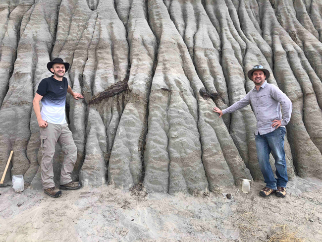
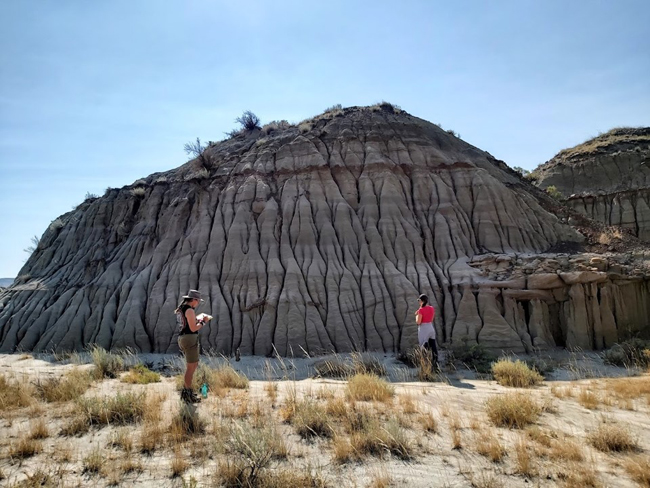
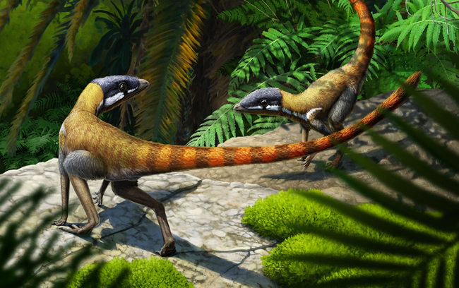
Research into a beautifully preserved Edmontosaurus fossil suggests that dinosaur mummies might be more common than previously thought. The Edmontosaurus specimen found by Tyler Lyson when exploring Slope County (North Dakota) and Hell Creek Formation exposures contained therein is providing palaeontologists with an insight into the fossilisation process that might produce a “dinosaur mummy”.

A mummified dinosaur was thought to require two mutually exclusive taphonomic processes in order to form. Firstly, to have the carcase exposed on the surface for a considerable portion of time to permit the remains to dry out and become desiccated. Secondly, rapid burial and deposition to preserve what remains of the corpse.

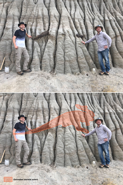
The taphonomy of the Edmontosaurus specimen (NDGS 2000), suggests that there may be other circumstances the lead to the mummified remains of dinosaurs.

Edmontosaurus "Dakota" Skin Preservation
Distribution and current state of preparation of the preserved skin on the Edmontosaurus specimen (NDGS 2000). Life reconstruction by Natee Puttapipat. Black areas in the diagram indicate portions of the skeleton apparently absent from the specimen, light grey areas indicate regions where the skeleton is preserved but no skin is currently preserved, red areas indicate regions where skin is present and is still undergoing preparation. The yellow shading indicate areas where the skin is fully prepared and were examined in this study. Picture credit: Drumheller et al (PLoS One).

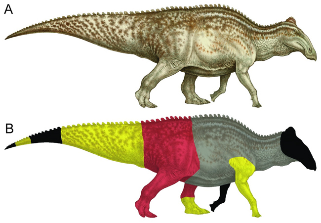
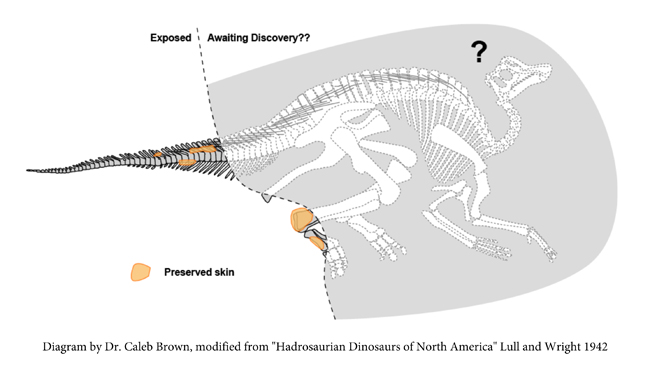
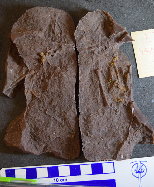
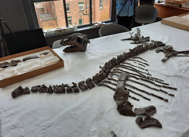
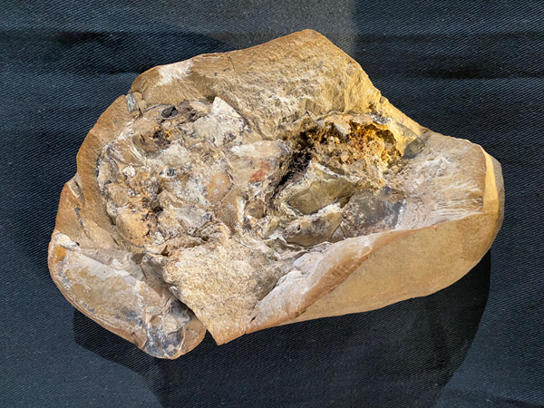
Dinosaur Mummies – Hooves and Fingers (E. annectens)

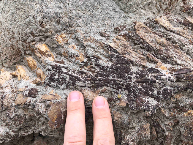
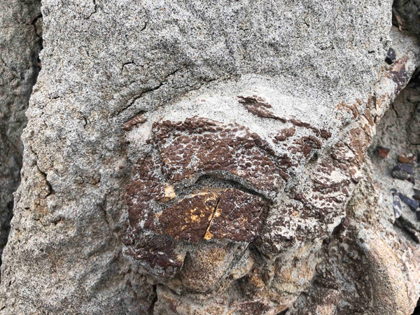
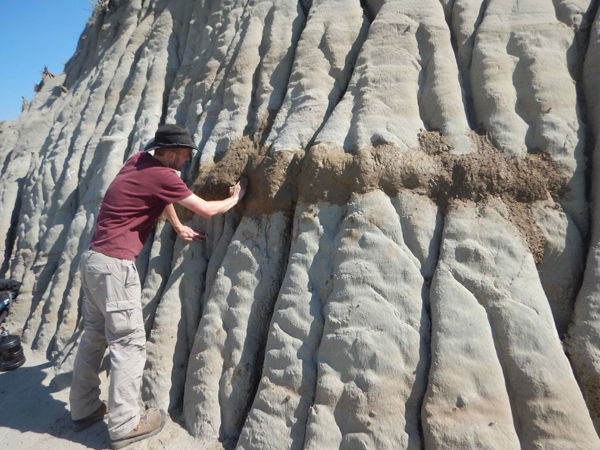
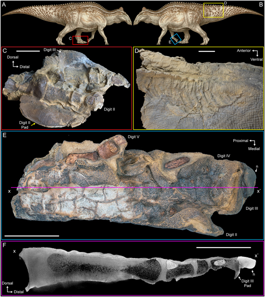
A team of scientists, including researchers from University of Tennessee–Knoxville, Knoxville, Tennessee and the North Dakota Geological Survey team, writing in the academic journal PLoS One propose a new explanation for how such fossil specimens might form. Large areas of desiccated and seemingly deflated skin have been preserved on the limbs and tail. Such is the degree of preservation of the front limb, (manus) that palaeontologists have discovered that Edmontosaurus (E. annectens) had a hoof-like nail on the third digit.

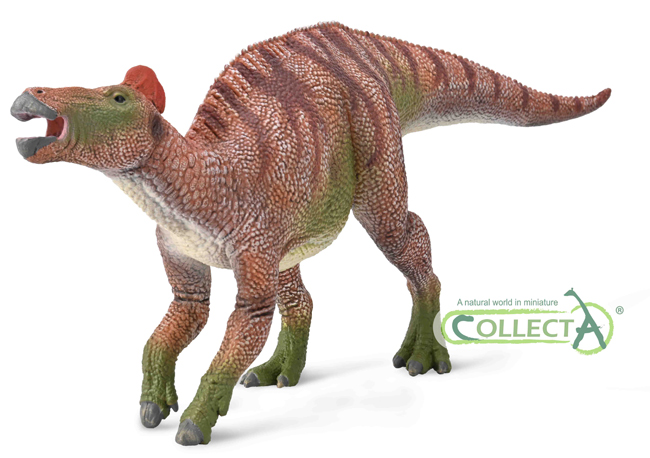
This discovery led to a substantial revision of Edmontosaurus limb anatomy in prehistoric animal replicas, as epitomised by the recently introduced CollectA Deluxe 1:40 scale Edmontosaurus.

CollectA Deluxe 1:40 scale Edmontosaurus dinosaur model
The new for 2022 CollectA Deluxe 1:40 scale Edmontosaurus dinosaur model. CollectA had wanted to introduce a replica of this iconic Late Cretaceous hadrosaur for some time. Many of the details such as the hoof-like third digit on the hand and the enlarged scales on the neck replicate what the scientists have discovered by studying the Edmontosaurus dinosaur mummy known as Dakota.

To read a blog article that contains a video review of the Edmontosaurus and explains more about the “dinosaur mummy” research: Everything Dinosaur Reviews the CollectA Deluxe Edmontosaurus Dinosaur Model.

Evidence of Scavenging

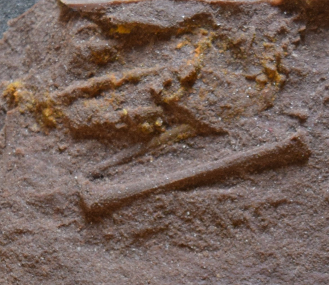
The research team identified bite marks from carnivores upon the dinosaur’s skin. These are the first examples of unhealed carnivore damage on fossil dinosaur skin, and furthermore, this is evidence that the dinosaur carcass was not protected from scavengers by being rapidly buried, yet it became a mummy nonetheless.

Many of the marks suggest bites from the conical teeth of crocodyliforms, although pathology associated with the tail is more difficult to interpret. The researchers suggest that some of the “V-shaped” patterns identified suggest that flexible, clawed digits rather than more rigidly fixed teeth, may have been responsible for these injuries. Perhaps these marks were caused by feeding deinonychosaurs (Dakotaraptor steini) or perhaps a juvenile T. rex.

Examining the Decomposition of Carcases

If the carcase was scavenged, then it was not buried rapidly and one of the supposed pre-requisites for “dinosaur mummification” did not occur with this fossil specimen. Instead, the researchers propose an alternative route for the creation of such remarkable fossils, a theory that has been influenced by what is observed in the world today. When scavengers feed on a carcase, they rip open the body and feed on the internal organs. Punctures made in the body allow fluids and gases formed by decomposition to escape, thus permitting the skin to dry out, forming a desiccated, dried out husk.

Evidence of desiccation of the Edmontosaurus fossil
Evidence of desiccation in the preserved remains of the Edmontosaurus (specimen number NDGS 2000). Note all scale bars equal 10 cm. Life reconstruction by Natee Puttapipat. Picture credit: Drumheller et al (PLoS One).

Dinosaur Skin More Commonly Preserved

The research team postulate that if the more durable soft tissues can persist some months prior to burial to permit desiccation to occur, then dinosaur skin fossils, although rare, are possibly, more commonly preserved than expected.

Edmontosaurus "Dakota" right manus preservation pathway
Cross sectional views through the right manus of the Edmontosaurus fossil (NDGS 2000). NDGS 2000 reconstruction in right lateral view (A). Right manus in dorsal view indicating the positions of the three cross sectional views (B). CT image along line x to x’ (C). CT image along line y to y’ (D). CT image along line z to z’ (E). In (C), (D), and (E), slice numbers from the original CT data are provided above each image. Paleoart in (A) by Natee Puttapipat. Scale bars equal 1 cm. Picture credit: Drumheller et al (PLoS One).

A New Theory on How “Dinosaur Mummies” Could Form

It is important to make clear that what a palaeontologist refers to as a “dinosaur mummy” is not the same as the mummified remains of an Egyptian deity. The skin and other soft tissues are permineralised, they are rock, although it is noted that molecular sampling of this Edmontosaurus specimen yielded putative dinosaurian biomarkers such as evidence of degraded proteins, suggesting that soft tissue was preserved directly in this specimen.

Generally, the two presumed prerequisites for mummification, that of being exposed on the surface for some time to permit the corpse to desiccate and rapid burial are incompatible. So, the researchers propose a new theory on how a “dinosaur mummy” could form:

  • A corpse is scavenged creating puncture marks to allow fluids and gases to escape.
  • Smaller organisms such as invertebrates and microbes exploit these punctures to access the internal organs and other parts of the skeleton.
  • Consumption from within in conjunction with decomposition allows the skin to deflate and to drape over the underlying bones that are more resistant to feeding and decay.
Edmontosaurus soft tissue preservation pathway.
Proposed soft tissue preservational pathway for the Edmontosaurus fossil. Incomplete predation and/or scavenging of the carcass creates openings in the body wall through which fluids and gasses can escape (A). Invertebrates and microbes (B) use those openings to access the internal tissues. Removal of internal soft tissues and drainage of fluids and gasses associated with decomposition allows the deflated skin and other dermal tissues to desiccate and drape over the underlying bones (C). Illustration by Becky Barnes. Picture credit: Drumheller et al (PLoS One).

The scientists hope that this new paper will help with the excavation, collection and preparation of fossils. The presence of soft tissues and biomarkers such as degraded proteins demonstrate that rapid burial may not be a pre-requisite to permit their preservation. As a result, such evidence as skin, soft tissue and biomarkers may be more common in the fossil record than previously thought.

The scientific paper: “Biostratinomic alterations of an Edmontosaurus “mummy” reveal a pathway for soft tissue preservation without invoking ‘exceptional conditions'” by Stephanie K. Drumheller, Clint A. Boyd, Becky M. S. Barnes and Mindy L. Householder published in PLoS One.

12 10, 2022

New Study into Stegoceras Muscles

By |2024-03-09T19:10:12+00:00October 12th, 2022|Categories: Adobe CS5, Dinosaur and Prehistoric Animal News Stories, Dinosaur Fans, Main Page, Palaeontological articles|0 Comments

Researchers have examined the musculature of a bone-headed dinosaur in a bid to better understand hypothesised intraspecific, head-butting combat. Pachycephalosaurs are a group of Late Cretaceous, bipedal ornithischian dinosaurs known from Asia and North America. They are characterised by their thickened skulls, which are sometimes adorned with lumps, bumps and spikes.

The skulls, some of which can be up to 20 cm thick have been the focus of a lot of research. It has been suggested that these thickened skull domes evolved as these dinosaurs indulged in intraspecific head-butting contests, either head-to-head impacts or using their heads to butt the flanks of their opponents.

A replica skull of Pachycephalosaurus wyomingensis.
Pachycephalosaurus wyomingensis replica skull. The thickened skull domes, which in some specimens of P. wyomingensis were up to 20 cm thick are thought to have evolved as these dinosaurs participated in intraspecific head-butting combat. Picture credit: Everything Dinosaur.

To read a blog post from 2011 looking at the evidence for head-butting combat in pachycephalosaurs: Study Supports Theory of Pachycephalosaur Intraspecific Combat (Head-butting).

Stegoceras Muscle Study

Writing in the open-access, on-line journal PLoS One, researchers from Carleton University, Ottawa in collaboration with Professor Phil Currie (University of Alberta) have examined the postcranial skeleton of a specimen of the pachycephalosaur Stegoceras validum to gain a better understanding of the musculature of the limbs, hips and the base of the tail. The specimen (UALVP 2) is one of the best preserved pachycephalosaur postcranial skeletons discovered to date and the best preserved pachycephalosaur discovered in Canada. The limb bones preserve muscle scars and other surface textures which enabled the research team to accurately construct the muscles associated with the forelimbs, hindlimbs and the pelvic region.

Pachycepahlosaurus indulging in head-butting combat.  Reporting upon a Stegoceras muscle study.
A charging Pachycephalosaurus.

Focusing on Muscles Not Bones

Unlike most studies relating to the Dinosauria, the fossil bones were not the central focus of this research. The scientists who include Professor Phil Currie (University of Alberta) and PhD student Bryan Moore (Carleton University), examined the bones to determine the layout, shape and size of the muscles that were attached to them. The team were interested in mapping the *myology of the back end of a pachycephalosaur so that they could assess how the postcranial skeleton would have assisted with the hypothesised head-butting contests.

The term *myology refers to the study of the shape, structure and arrangement of muscles.

Stegoceras muscle study.
Pelvic and hind limb muscular reconstruction of Stegoceras validum. Superficial musculature in lateral view (A). Deep musculature in lateral view (B). Superficial musculature in anterodorsolateral view (C) and (D) deep musculature in anterodorsolateral view. Picture credit: Moore et al/PLoS One.

Strong Legs and a Wide Pelvis

The study of specimen number UALVP 2 demonstrated that the forelimbs of Stegoceras validum were not especially robust and strong, particularly in comparison to early, lizard-hipped bipeds such as the Triassic theropod Tawa hallae. However, in contrast, in Stegoceras the hind limbs and pelvic area were more robust with large, powerful muscles associated with the pelvis, the thighs and the base of the tail. These larger muscles, in combination with the wide pelvis and stout hind limbs, produced a stronger, more stable pelvic structure that would have proved advantageous during hypothesised intraspecific head-butting contests.

The new for 2020 Wild Safari Prehistoric World Pachycephalosaurus dinosaur model.
The new for 2020 Wild Safari Prehistoric World Pachycephalosaurus model. The model has a large dent in its skull dome, suggesting damage resulting from a headbutting contest with a rival.

The picture above shows a Pachycephalosaurus dinosaur model from the Wild Safari Prehistoric World range, to view this range of figures in stock at Everything Dinosaur: Safari Ltd Dinosaur Models.

The research team concludes that the hind quarters of Stegoceras evolved to help this small dinosaur deliver and absorb impact forces associated with the proposed head-butting behaviour. The scientists suggest that more research is needed to examine the potential velocity at which the thickened skull could be propelled forward during such contests. They propose additional research assessing the postcranial properties of other pachycephalosaurs and comparing their bauplan with similar sized dinosaurs such as Thescelosaurus (T. neglectus).

The scientific paper: “The appendicular myology of Stegoceras validum (Ornithischia: Pachycephalosauridae) and implications for the head-butting hypothesis” by Bryan R. S. Moore, Mathew J. Roloson, Philip J. Currie, Michael J. Ryan, R. Timothy Patterson and Jordan C. Mallon published in PLoS One.

The Everything Dinosaur website: Everything Dinosaur.

6 10, 2022

Spotting A Rare Hadrosaur Fossil

By |2024-03-19T16:47:25+00:00October 6th, 2022|Categories: Dinosaur and Prehistoric Animal News Stories, Dinosaur Fans, Main Page, Palaeontological articles, Photos/Pictures of Fossils|0 Comments

Sometimes serendipity and palaeontology combine, for example, a sharp-eyed field team member spotting a hadrosaur fossil specimen eroding out of a small hill in the Dinosaur Provincial Park (Alberta, Canada). The fossils could represent a rare skeleton of a juvenile and there is evidence that skin impressions have been preserved.

Whilst hadrosaur fossils are relatively common in this part of southern Alberta, the animal’s tail and right hind foot are orientated in the hillside to suggest that the entire skeleton may still be preserved within the rapidly eroding mudstone.

Standing next to the exposed hadrosaur skeleton.
Brian Pickles (left) and Caleb Brown (right) stand next to the exposed skeleton. Picture credit: Melissa Dergousoff/University of Reading.

Potentially a Very Significant Fossil Discovery

Whole dinosaur skeletons are extremely rare, this specimen tentatively referred to as a “dinosaur mummy” could provide important new information on juvenile hadrosaurs and the ontogeny of duck-billed dinosaurs.

Diagram of potential hadrosaur specimen
A diagram of the potential hadrosaur skeleton showing exposed parts with skin impressions and the potential orientation of the rest of the skeleton. Picture credit: Caleb Brown.

Spotting a Hadrosaur

The exposed caudal vertebrae (tail bones) show preserved skin impressions as does the exposed right ankle. The size of the bones and the distance between the tail and the astragalus (ankle) suggest that these are the fossilised remains of a young hadrosaur.

Close-up view of the exposed caudal vertebrae with preserved skin impressions.
A close-up view of the exposed caudal vertebrae with preserved skin impressions. Picture credit: Royal Tyrrell Museum of Palaeontology.
View of the exposed ankle bones with skin impressions.
A view of the exposed ankle bones with skin impressions. Picture credit: Royal Tyrrell Museum of Palaeontology.

Discovering a Duck-billed Dinosaur

During a field school scouting visit in 2021 to look for possible excavation sites, Dr Brian Pickles (University of Reading) was leading a small team examining one location when volunteer crew member Teri Kaskie spotted the fossil skeleton protruding from the hillside.

The "Hadrosaur Hill"
Teri Kaskie (right) and (left) Melissa Dergousoff stand next to the hill containing the hadrosaur skeleton. Picture credit: Brian Pickles University of Reading.

The first international palaeontology field school is taking place, involving academics and students from the University of Reading and the University of New England in Australia. In collaboration with researchers from the Royal Tyrrell Museum (Drumheller, Alberta), the team are working together to excavate the skeleton and ensure the material that remains in the hill is protected from the elements.

The first part of the conservation work involves coating the fossil site in a thick layer of mud, to help conserve the delicate fossils and to prevent erosion.

Covering the exposed fossils with mud
Covering the exposed fossils with mud to provide protection. Picture credit: Royal Tyrrell Museum of Palaeontology.

An Exciting Fossil Discovery

Commenting on the significance of this hadrosaur fossil find, Dr Pickles stated:

“This is a very exciting discovery, and we hope to complete the excavation over the next two field seasons. Based on the small size of the tail and foot, this is likely to be a juvenile. Although adult duck-billed dinosaurs are well represented in the fossil record, younger animals are far less common. This means the find could help palaeontologists to understand how hadrosaurs grew and developed.”

Vertebrate palaeontologist from the Royal Tyrrell Museum, Dr Caleb Brown added:

“Hadrosaur fossils are relatively common in this part of the world but another thing that makes this find unique is the fact that large areas of the exposed skeleton are covered in fossilised skin. This suggests that there may be even more preserved skin within the rock, which can give us further insight into what the hadrosaur looked like.”

Protecting the exposed hadrosaur fossils
The burlap screen erected over the exposed fossils to help protect the material from erosion. Picture credit: Royal Tyrrell Museum of Palaeontology.

A Substantial Project

Collecting the entire skeleton is going to take many months and the site will have to be closed down and secured as the weather worsens towards winter. It may take several field seasons to complete this work. Once the specimen has been removed from the field, it will be delivered to the Royal Tyrrell Museum’s Preparation Laboratory, where skilled technicians will work to uncover and conserve the fossilised bones.

At this time, the scientists are unsure as to how complete the specimen is and which genus the fossils represent. Species identification will only be possible if a substantial proportion of the skeleton, including skull material can be recovered.

Exposed hadrosaur skeletal material in the Dinosaur Provincial Park Formation
Brian Pickles and Caleb Brown stand next to the exposed skeleton with an illustration showing estimated skeleton size and potential position. Picture credit: Melissa Dergousoff University of Reading with diagram by Caleb Brown.

Which Hadrosaur?

Several different types of hadrosaur are known from the Dinosaur Provincial Park Formation (Campanian faunal stage). Lambeosaurines are represented by Corythosaurus, Parasaurolophus and Lambeosaurus whilst members of the Saurolophinae subfamily represented include Gryposaurus and Prosaurolophus. As more of the skeleton is prepared, the researchers are hopeful that they will be able to confirm the species.

Everything Dinosaur acknowledges the assistance of a media release from Reading University in the compilation of this article.

5 10, 2022

Unravelling the Ancestry of the Pterosauria

By |2022-10-05T16:05:15+01:00October 5th, 2022|Categories: Dinosaur and Prehistoric Animal News Stories, Dinosaur Fans, Main Page, Palaeontological articles, Photos/Pictures of Fossils|0 Comments

A fossil specimen found in Scotland more than 100 years ago is helping to unravel the ancestry of the Pterosauria. A new study of tiny and difficult to interpret fossils representing a reptile named Scleromochlus taylori has provided palaeontologists with a fresh perspective on the evolution of the pterosaurs.

The research, published in the academic journal “Nature”, was undertaken by scientists led by Dr Davide Foffa, a Research Associate at National Museums Scotland, and now a Research Fellow at the University of Birmingham. The study, which involved analysis of the fossils using Computed Tomography (CT scans), has enabled the first, accurate skeletal reconstruction of Scleromochlus taylori.

An early ancestor of the Pterosauria
A tiny reptile measuring approximately 20 cm in length, Scleromochlus is now thought to be an early ancestor of the Pterosauria. Picture credit: Gabriel Ugueto.

Anatomical Details Reveal Link with the Pterosauromorpha

The CT scans revealed new anatomical details that conclusively identify the Scleromochlus genus as a close pterosaur relative. Phylogenetic assessment places this small, agile reptile within a group known as Pterosauromorpha. The Pterosauromorpha comprises pterosaurs and their close relatives the lagerpetid reptiles.

Scleromochlus fossil casts (ancestry of the Pterosauria).
Scleromochlus fossil Casts (NHMUK-PV-R3557). Casts created enabled the scientists to reconstruct the skeleton. Picture credit: London Natural History Museum/University of Birmingham.

Identifying the Ancestry of the Pterosauria (Lagerpetonidae)

Geographically widespread in the Late Triassic, the Lagerpetonidae were typically small and fleet-footed reptiles, classified as basal avemetatarsalians, the branch of the Archosauria leading to birds, dinosaurs and the Pterosauria. Previously thought to be close to the evolutionary tree of the Dinosauria, more recent research, including this newly published paper suggests that the lagerpetids were closer to the pterosaurs (members of the Pterosauromorpha).

Most lagerpetids are described as being about the size of cat or small dog, however, Scleromochlus was smaller, with an estimated body length of around 20 cm.

This new study supports the hypothesis that the first flying reptiles (pterosaurs) evolved from small, likely bipedal ancestors. The placement of the lagerpetids within the avemetatarsalians had caused extensive debate. This paper argues that Scleromochlus, represented an evolutionary step in the direction of pterosaurs.

Poorly Preserved Fossils – the Elgin Reptiles

Analysis of the Scleromochlus fossil material using more traditional methods is extremely difficult. The non-destructive CT scans enabled the research team to examine the fossilised bones in exquisite detail and revealed new anatomical details that had not been observed before.

The sandstone block containing the bones come from Morayshire in north-eastern Scotland, near to the town Elgin. Collectively fossils of vertebrates from these deposits are known as the Elgin Reptiles. The fossils are held mostly in the collections of National Museums Scotland, Elgin Museum and the Natural History Museum. The latter holds Scleromochlus, which was originally found at Lossiemouth.

Scleromochlus fossil (ancestry of the Pterosauria).
A Scleromochlus fossil. Picture credit: Professor Paul Barrett London Natural History Museum/University of Birmingham.

Ancestry of the Pterosauria

Commenting on the significance of the research, Dr Foffa stated:

“It’s exciting to be able to resolve a debate that’s been going on for over a century, but it is far more amazing to be able to see and understand an animal which lived 230 million years ago and its relationship with the first animals ever to have flown. This is another discovery which highlights Scotland’s important place in the global fossil record, and also the importance of museum collections that preserve such specimens, allowing us to use new techniques and technologies to continue to learn from them long after their discovery.”

Professor Paul Barrett at the Natural History Museum added:

“The Elgin reptiles aren’t preserved as the pristine, complete skeletons that we often see in museum displays. They’re mainly represented by natural moulds of their bone in sandstone and – until fairly recently – the only way to study them was to use wax or latex to fill these moulds and make casts of the bones that once occupied them. However, the use of CT scanning has revolutionized the study of these difficult specimens and has enabled us to produce far more detailed, accurate and useful reconstructions of these animals from our deep past.

Scleromochlus fossil casts (close view)
A close-up view of a cast of NHMUK-PV-R3557 with clearly defined Scleromochus fossil bones. Picture credit: London Natural History Museum/University of Birmingham.

The First Vertebrates to Evolve Powered Flight

Co-author of the scientific paper, professor Sterling Nesbitt (Virgina Tech) commented:

“Pterosaurs were the first vertebrates to evolve powered flight and for nearly two centuries, we did not know their closest relatives. Now we can start filling in their evolutionary history with the discovery of tiny close relatives that enhance our knowledge about how they lived and where they came from.”

Everything Dinosaur acknowledges the assistance of a media release from the University of Birmingham in the compilation of this article.

The scientific paper: “Scleromochlus and the early evolution of the Pterosauromorpha” by Davide Foffa, Emma M. Dunne, Sterling J. Nesbitt, Richard J. Butler, Nicholas C. Fraser, Stephen L. Brusatte, Alexander Farnsworth, Daniel J. Lunt, Paul J. Valdes, Stig Walsh and Paul M. Barrett published in Nature.”

1 10, 2022

Classifying Palaeontology in Video Games

By |2022-10-02T07:14:45+01:00October 1st, 2022|Categories: Dinosaur and Prehistoric Animal News Stories, Dinosaur Fans, Main Page, Palaeontological articles|4 Comments

Everything Dinosaur team members wrote a blog post a few days ago about how palaeontology is depicted in video games. The University of Birmingham led research, published recently in the journal EGUsphere Geoscience Communications, examined how the science of palaeontology is portrayed to the public, at a time when many people get a lot of their knowledge from media and entertainment.

To read Everything Dinosaur’s blog post about this research: Palaeontology in Video Games.

Nanmu Studio Indominus Berserker Rex
A model of a genetically designed dinosaur. Inspired by InGen? Video games and other media such as films are having a big influence on how the Dinosauria are perceived by the public.

Classifying How Video Games Depict Palaeontology

In order to understand how palaeontology and other related Earth sciences are portrayed, the research team looked at palaeontological content and classified it into a number of categories.

Here is the list of categories the team used:

  • Ancient animals as adversaries – using ancient animals – typically dinosaurs and pterosaurs – as adversaries that must be killed. Examples include The Legend of Zelda, Tomb Raider and Peter Jackson’s King Kong video game.
  • Ancient animals as tools – for example Yoshi, an omnivorous theropod dinosaur who first appeared in Super Mario World as a mount for the Mario brothers in their adventures to rescue Princess Peach.
  • Fossils as collectables – items found throughout the game setting to help the player progress through the game. For example, Red Dead Redemption 2 features a side quest to locate 30 fossil specimens in order to assist an aspiring palaeontologist.
  • Ancient animal management simulators, otherwise referred to as “park sims” – games such as the Jurassic World Evolution aim to construct a financially viable park or zoo.
  • Ancient animal simulators – players control an individual animal through an entire life cycle in a natural, open world, environment such as Saurian or Ancestors: The Humankind Odyssey.

Helping or Hindering the Perception of Palaeontology

Once the researchers had classified the video game content, they set about devising a rating system to determine whether the depiction hindered or helped the science of palaeontology and how it was depicted in cyberspace compared to the real world.

The factors analysed to determine the impact were:

  • Ancient death machines – ancient animals as vicious, frightening and brutish enemies for players to combat. AKA ‘monsterification’, the animal’s proportions and features like claws and horns are exaggerated – for example in Ark: Survival Evolved.
  • Fossil = dinosaur = Tyrannosaurus: lack of palaeodiversity – Dinosaurs have become almost synonymous with ancient life in the entertainment industry. Ancient animal diversity is often limited to a handful of “iconic” species recycled by the entertainment industry.
  • Palaeotrivia – In order to help players understand complex topics or introduce story elements, many games feature in-built encyclopaedias about characters, objects and locations in the game setting – particularly in park management games such as Jurassic World: Evolution series.
  • Depiction of palaeontological science – most palaeo-video games integrate science into gameplay that increases the knowledge of the player, but they can take creative liberties in order to make engaging gameplay mechanics. In essence, these games may depart from the science narrative all too readily.
  • Representation of ethics in palaeontological video games – Palaeontology has a long colonial history with deep-rooted exploitative practises which appear within games unchallenged, for example the illegal buying and selling of fossils in Jurassic World: Evolution.
  • Male, pale and stale – palaeontologists in video games are typically depicted as old, white men or the ‘Indiana Jones stereotype’ – for example Stardew Valley and Dinosaur Fossil Hunter.
  • Perpetuation of harmful, misogynistic, and racist tropes – while female lead characters are becoming more commonplace, games still sexualise women more than their male counterparts, particularly in depictions of early humans.

The Most Common Palaeontological Tropes

By highlighting the most common palaeontological tropes, both negative and positive, identified in video games, the researchers hope to assist science communicators when working in the virtual reality or gaming sector. In addition, by flagging common misconceptions and harmful tropes, the research team aims to raise awareness amongst game developers, who may be unaware that they could be perpetuating negative notions about the Earth sciences.

Mosasaurus model.
Nanmu Studio Mosasaurus “Lord of the Abyss”. A marine reptile model inspired by how prehistoric animals are depicted in movies and video games.

The prehistoric animal images used to illustrate this article feature Nanmu Studio prehistoric animal models. To view the Nanmu Studio range in stock at Everything Dinosaur: Nanmu Studio Dinosaur and Prehistoric Animal Models.

The scientific paper: “The perception of palaeontology in commercial off-the-shelf video games and an assessment of their potential as educational tools” by Thomas Clements, Jake Atterby, Terri Cleary, Richard P. Dearden and Valentina Rossi published in EGUsphere Geoscience Communications.

28 09, 2022

Research Suggests Ancient “Sharks” Appeared Much Earlier

By |2024-03-19T16:49:26+00:00September 28th, 2022|Categories: Dinosaur and Prehistoric Animal News Stories, Main Page, Palaeontological articles, Photos/Pictures of Fossils|0 Comments

Newly published research suggests that ancient “sharks” appeared much earlier than previously thought. A fossil from China represents a new species of jawed fish (Qianodus duplicis) and its discovery suggests that fishes with true jaws first evolved in the Early Silurian.

Qianodus duplicis life reconstruction
Qianodus duplicis, a new genus and species of an early Silurian gnathostome. Picture credit: Heming Zhang/University of Birmingham.

An Early Silurian Origin of Shark-like Jaws

The scientific paper, published in the journal “Nature” identifies Q. duplicis as the earliest record of a toothed gnathostome known to science. Its discovery extends the record of toothed gnathostomes by some 14 million years from the Late Silurian into the Early Silurian (around 439 million years ago).

The fossils (a handful of tiny teeth), found in China represent the earliest direct evidence for jawed vertebrates known to science.

Previously, the earliest jawed fish to be positively identified, included species from the Late Silurian, fossils thought to date from around 424 million years ago. These include the placoderms (Class Placodermi) partially armoured gnathostomes, and sarcopterygians, bony “lobe-finned” fishes found initially in China and Vietnam.

Confirming Evidence from Fossil Fish Scales

Co-author of the paper, Dr Ivan Sansom (University of Birmingham), commented:

“Until this point, we’ve picked up hints from fossil scales that the evolution of jawed fish occurred much earlier in the fossil record, but have not uncovered anything definite in the form of fossil teeth or fin spines.”

Construction workers building a new road in Guizhou Province uncovered fossil material and field teams from the Chinese Institute of Vertebrate Palaeontology and Palaeoanthropology (IVPP) and the Qujing Normal University (QJNU), were despatched to take samples and to analyse the Silurian-aged deposits.

The scientists found numerous scales, but also recovered several miniscule fossil teeth between 1.5 mm and 2.5 mm in length.

Dr Sansom explained:

“Scales are relatively easy to find because they are so plentiful, but teeth are much scarcer. The scale and speed at which IVPP and QJNU colleagues were able to sift through the material enabled us to identify these scarce remains more effectively than in previous projects.”

Around twenty of the tiny fossil teeth turned out to be from the same species (Qianodus duplicis). From the arrangement of the teeth and their morphology, the team established that they would have come from a fish with an arched jaw margin, with offset tooth rows, similar to those found in extant sharks. The team used a range of techniques, including Computed Tomography (CT scans), to establish a date for the samples.

What’s in a Name?

The new species Qianodus duplicis comes from “Qian” the ancient name for Guizhou Province, “odus” from the Greek for tooth, and duplicis, or double, referring to the paired rows of teeth.

A Cartilaginous Fish – Fanjingshania renovata

In a separate paper, also published today in Nature, the team also identified fossil elements that relate to “fin spines”, bony projections in front of the fins which can be seen today on Port Jackson sharks. These spiny structures form the basis for the identification of a new species Fanjingshania renovata named after Mount Fanjingshan which is close to the locality from where the fossil material was collected. The species name “renovata”, acknowledges renewal, the remodelling of the base of the spines and scales.

Fanjingshania renovata life reconstruction.
Fanjingshania renovata life reconstruction. Picture credit: Heming Zhang/University of Birmingham.

Lead author of both papers Dr Plamen Andreev (Qujing Normal University), commented:

“The early so-called “spiny sharks” had these features on all of their fins, but the examples that we have found belong to a much earlier period. These are the first creatures that we would recognise today as fish-like, evolving from creatures often referred to as “clams with tails”, from earlier in the Ordovician period.”

Ancient “Sharks” Appeared Much Earlier

Cartilaginous fish (chondrichthyans), including sharks, separated off at some point from osteichthyans (bony fish and tetrapods), from which our own species eventually evolved. The point at which this occurred, however, is obscured within ghost lineages in the Ordovician, where only hints in the fossil record have been found. Precisely how and when this separation happened, therefore, remains ambiguous.

Ancient "sharks" appeared much earlier.
Fanjingshania life reconstruction in lateral view. Picture credit: Heming Zhang/University of Birmingham.

Everything Dinosaur acknowledges the assistance of a media release from the University of Birmingham in the compilation of this article.

The scientific paper: “The oldest gnathostome teeth” by Plamen S. Andreev, Ivan J. Sansom, Qiang Li, Wenjin Zhao, Jianhua Wang, Chun-Chieh Wang, Lijian Peng, Liantao Jia, Tuo Qiao and Min Zhu published in Nature.

The scientific paper announcing Fanjingshania renovata: “Spiny chondrichthyan from the lower Silurian of South China” by Plamen S. Andreev, Ivan J. Sansom, Qiang Li, Wenjin Zhao, Jianhua Wang, Chun-Chieh Wang, Lijian Peng, Liantao Jia, Tuo Qiao and Min Zhu published in Nature.

27 09, 2022

Palaeontology in Video Games

By |2023-02-07T09:25:47+00:00September 27th, 2022|Categories: Adobe CS5, Dinosaur and Prehistoric Animal News Stories, Dinosaur Fans, Main Page, Palaeontological articles|3 Comments

Palaeontology, dinosaurs and prehistoric animals are frequently subjects for video games. Players have the opportunity to construct their own “Jurassic Park”, go fossil collecting and combat dinosaurs. A new study indicates that these games may contain negative and harmful themes that can confuse and lead to misconceptions about palaeontology.

An international team of researchers, led by scientists from the University of Birmingham, played and studied a variety of video games containing elements of palaeontology. An eclectic range of video games were sampled including Super Mario World, Animal Crossing and the “Jurassic Park/World” games. The team attempted to map what was “palaeo-fact” and what was “palaeo-fiction”.

A timeline of palaeontology in video games
Timeline of palaeontology in video games. Picture credit: Clements et al/University of Birmingham.

Sorting “Palaeo-fact” from “Palaeo-fiction”

Dr Thomas Clements (University of Birmingham) and a co-author of the study explained:

“Loads of people are inspired by and get their understanding of dinosaurs from movie blockbusters like Jurassic Park, but no one talks about how massive the gaming industry is in shaping not only the public’s understanding of ancient life and also of paleontological science.”

A Triassic forest from a video game.
Gamers will be able to explore a number of terrestrial environments. A Triassic forest environment from a video game. Picture credit: Daniel Carter.

Pleasantly Surprised by the Accuracy

The researchers were pleasantly surprised by the accuracy of some of the games. However, they quickly identified numerous negative tones that were repeatedly reflected in the gaming environment.

Dr Clements commented:

“When we played through many of these games, we were pleasantly surprised about the accuracy of games like Animal Crossing that provide accurate and educational information in a fun and engaging way. However, we also found that many games contain misleading, negative, and sometimes quite damaging themes – many already widespread issues in the gaming industry. It is common for palaeo-games to contain ethically dubious science, the illegal collection of fossils, ‘monsterification’ of animals, poor representation of minority groups, and the hypersexualisation of women.”

Writing in the journal EGUsphere Geoscience Communications, the scientists analysed the representation of palaeontology in hundreds of video games, classifying them into several categories. They then defined a number of factors which may help or hinder a video game’s effectiveness in promoting palaeontology to a wider audience. Their study has implications for ways that science communicators can address these issues when talking to the public about palaeontology and has a wider role in helping to support gender equality and ethnic diversity.

Portraying Palaeontology to the Public

Co-author Jake Atterby (University of Birmingham), stated:

“This paper is about how the science of palaeontology is portrayed to the public, at a time when many people get a lot of their knowledge from media and entertainment. Audiences can subconsciously learn from the media they consume, including depictions of our science that are deliberately exaggerated for entertainment. This can give players a false impression of ancient life and the work that we do. It is important for palaeontologists to understand the public’s perception of our science to help when we communicate our research.

Everything Dinosaur acknowledges the assistance of a media release from the University of Birmingham in the compilation of this article.

The scientific paper: “The perception of palaeontology in commercial off-the-shelf video games and an assessment of their potential as educational tools” by Thomas Clements, Jake Atterby, Terri Cleary, Richard P. Dearden and Valentina Rossi published in EGUsphere Geoscience Communications.

23 09, 2022

Allometric Growth – A Helpful Explanation

By |2024-04-13T08:54:47+01:00September 23rd, 2022|Categories: Dinosaur Fans, Key Stage 3/4, Main Page, Palaeontological articles, Teaching|0 Comments

We received an email earlier this week with an enquiry about the term allometric growth which had been found in a scientific paper the emailer had been reading. Our emailer wrote to ask what did this term mean?

Allometric growth is a term used to describe the growth of an organism whereby different parts develop at different rates. The appearance of the organism will change as it grows and matures.

Papo Triceratops and baby Triceratops models - allometric growth.
The Papo baby Triceratops figure next to the adult Papo Triceratops. Picture credit: Everything Dinosaur.

Triceratops Allometric Growth

In the picture (above) a model of a juvenile Triceratops is compared with a model of a fully-grown, mature Triceratops. In the juvenile the head is proportionately larger and the skull frill and horns are very different in their morphology compared to the adult. This is an example of allometric growth. Allometry is the study of body size relative to body shape, it is often partnered in scientific papers with ontogeny which is the term used to describe how organisms develop and grow.

The Chinese model-making company PNSO have recently introduced some replicas that demonstrate how dinosaurs changed as they grew and matured. For example, the company recently introduced (2022), a 1:35 scale replica of an adult chasmosaurine ceratopsian (Torosaurus latus) and a juvenile. The models, entitled Aubrey and Dabei were supplied with posters and a full-colour, illustrated booklet.

PNSO Aubrey and Dabei (Torosaurus dinosaur models)
The 1:35 scale PNSO Torosaurus models (Aubrey and Dabei). The adult Torosaurus figure has an articulated lower jaw. Aubrey is the name of the adult Torosaurus in this model set, it towers over the juvenile (Dabei). The pair of models illustrate the biological concept of allometric growth – how organisms often change shape as they develop as different parts of the animal grow at different rates. Picture credit: Everything Dinosaur.

To view the range of PNSO models and figures available from Everything Dinosaur: PNSO Age of Dinosaurs.

Back in 2019, Everything Dinosaur received an almost identical enquiry about how to define allometric growth. At the time, we wrote a short blog post highlighting our explanation.

To read our earlier article: Defining Allometric Growth.

22 09, 2022

Bring Back “April” Campaign Launched to Restore Unique Fossil Specimen

By |2024-04-13T08:57:39+01:00September 22nd, 2022|Categories: Adobe CS5, Dinosaur and Prehistoric Animal News Stories, Dinosaur Fans, Main Page, Palaeontological articles|0 Comments

The University of Manchester has launched a campaign to bring back “April” the Tenontosaurus. Manchester Museum is asking for public support to install a stunning Tenontosaurus fossilised skeleton, one of the most complete specimens of this Early Cretaceous ornithopod ever found.

The Museum is hoping to raise £10,000 GBP ($11,280.00 USD), to enable the Tenontosaurus to be restored and installed in the Dinosaurs and Fossils gallery as an exhibit with the 110-million-year-old fossil bones in their correct anatomical positions.

The fossilised remains of April the Tenontosaurus laid out in anatomical position.
The fossilised remains of “April” the Tenontosaurus partially laid out in anatomical position. Picture credit: The University of Manchester.

The Tenontosaurus specimen (MANCH LL.12275) was purchased by the University of Manchester in 1999. It has been the subject of a recent scientific paper which confirmed the presence of gastroliths (stomach stones), the first evidence of gastroliths to be identified in a derived member of the Ornithopoda.

To read about this scientific research: “April” the Tenontosaurus Reveals Her Secrets.

Bring Back “April”

The stunning fossil specimen comes from Montana, and it is affectionately named “April” after the wife of Barry James who originally prepared the fossil for display. When first put on show to the public it was portrayed in an upright position, like a super-sized kangaroo with its tail resting on the ground. However, research from Earth Sciences students from the University of Manchester has shed light on how the skeleton would have walked and posed in life.

Tenonotosaurus skeleton made ready for exhibition.
The original mount of “April” the Tenontosaurus. The skeleton was dismantled and put into storage when a replica of a T. rex skeleton made its debut at the museum. Picture credit: The University of Manchester.

Thousands of Hours of Restoration Work is Required

The Curator of the Earth Science Collections at the Manchester Museum David Gelsthorpe outlined the aims of the fund-raising effort and explained some of the problems that this restoration project will pose.

He commented:

“April is a Tenontosaurus purchased by Manchester Museum in 1999 and was previously displayed standing upright. Over the past few years, we have been working with a team of Earth Sciences students from the University of Manchester to carefully study April’s bones and find out more about her. Using their palaeontology skills and computer modelling, their research has helped us to better understand how she would have moved on all fours. As well as changing the way the skeleton stands, over 10,000 hours of careful conservation work is required to restore its bones.”

The Museum is requesting donations to help bring “April” back to her best and to permit her to be once again an integral part of the Museum’s Dinosaurs and Fossils Gallery.

To read more about this campaign and to make a donation: Support Manchester Museum Help Bring “April” Back.

The restored skull of "April" the Tenontosaurus
Tenontosaurus skull in left lateral view. The stunning and beautifully restored “April” the Tenontosaurus skull. Picture credit: The University of Manchester.

A New Dinosaur Display

If the fund raising succeeds than the Tenontosaurus specimen will form the focal point of a brand-new exhibition devoted entirely to the Dinosauria, planned for April 2023. Visitors will have the opportunity to view prehistoric giants, such as “Stan” the Tyrannosaurus rex cast and to learn about British dinosaur fossil discoveries. The demise of the Dinosauria and many other types of animals and plants as a result of a mass extinction event, some sixty-six million years ago, will be linked to today’s problems of climate change and the current rate of extinction.

Manchester Museum.
The front entrance of the Manchester Museum, a neo-Gothic building that houses approximately 4.5 million artefacts from the natural sciences and human history. A new dinosaur exhibit is scheduled to open in February 2023. Picture credit: The University of Manchester.

Plans for the new dinosaur exhibit are part of a larger scheme of improvements planned for Manchester Museum which has been entitled “Hello Future”.

To play your part and contribute to “April’s” restoration, please visit: Support Manchester Museum. Every donation will go towards helping to put “April” back on display.

Everything Dinosaur acknowledges the assistance of a media release from Manchester Museum in the compilation of this article.

16 09, 2022

A Devonian Heart

By |2023-02-25T20:51:42+00:00September 16th, 2022|Categories: Adobe CS5, Dinosaur and Prehistoric Animal News Stories, Main Page, Palaeontological articles, Photos/Pictures of Fossils|0 Comments

Researchers have discovered a 380-million-year-old fossilised heart, along with other internal organs of prehistoric fish. This exciting discovery is not only helping palaeontologists to understand the internal organs of placoderms, these fossils are also providing a fresh perspective on our own evolution.

The Gogo fish fossil where the 380-million-year-old, 3-D preserved heart was discovered by the research team.
The Gogo fish fossil where the 380-million-year-old, 3-D preserved heart was discovered by the research team. Picture credit: Curtin University.

Fossilised Internal Organs in Ancient Armoured, Jawed Fish

The remarkably well-preserved fossils representing the Devonian placoderm Compagopiscis, come from the famous Gogo Formation, in the Kimberley region of Western Australia. The Gogo Lagerstätte preserves the fauna that once thrived on an ancient, shallow water reef. The fossils are thought to be around 380 million years old (Frasnian faunal stage of the Late Devonian).

Gogo fish diorama
Gogo fish diorama at the West Australia Museum Boola Bardip depicting the typical fauna associated with the Gogo Lagerstätte. Typical inhabitants of a tropical reef that existed approximately 380 million years ago. Picture credit: Curtin University.

Soft Tissues Including Liver and Intestines but No Evidence of Lungs

The international research team led by scientists from Curtin University and the Western Australia Museum have published their findings in the academic journal “Science”. They have identified a number of internal organs preserved in three-dimensions including the liver and intestines. Additionally, the fossils show that the lungs are absent, refuting the hypothesis that lungs are ancestral in jawed vertebrates.

CT scans and interpretive models showing the heart of the placoderm Compagopiscis.
CT scans and interpretive models showing the heart of the placoderm Compagopiscis. Picture credit: Curtin University.

Details Revealed by Computerised Tomography

In collaboration with scientists at the Australian Nuclear Science and Technology Organisation in Sydney and the European Synchrotron Radiation Facility in Grenoble, France, researchers used neutron beams and synchrotron x-rays to scan the specimens, still embedded in limestone concretions. They constructed three-dimensional, computer-generated images of the soft tissues inside them based on the different densities of minerals deposited by the bacteria and the surrounding rock matrix.

Lead researcher and John Curtin Distinguished Professor Kate Trinajstic (Curtin’s School of Molecular
and Life Sciences and the Western Australian Museum), commented that to find three-dimensionally preserved tissues in Devonian fossils was an extremely rare event.

Professor Trinajstic explained:

“As a palaeontologist who has studied fossils for more than 20 years, I was truly amazed to find a 3-D and
beautifully preserved heart in a 380-million-year-old ancestor. Evolution is often thought of as a series of small steps, but these ancient fossils suggest there was a larger leap between jawless and jawed vertebrates. These fish literally have their hearts in their mouths and under their gills – just like sharks today.”

The Complex Heart of an Arthrodiran

This is the first time a 3-D representation of the heart of a member of the Arthrodira has been found. The Arthrodira are an extinct Order of jawed, armoured fish within the Class Placodermi. They thrived in the Devonian and some arthrodirans evolved into apex predators such as the huge Dunkleosteus.

Dunkleosteus on display.
The spectacular Dunkleosteus exhibit. A giant apex predator of the Devonian. Picture credit: Everything Dinosaur.

The surprising complex heart is s-shaped and consists of two chambers with the smaller chamber sitting on top. Professor Trinajstic stated that these features were advanced in such early vertebrates, offering scientists a unique perspective on how the head and neck region began to change to accommodate jaws, a critical stage in the evolution of animals with backbones.

Reconstruction of arthrodire internal anatomy.
Reconstruction of arthrodire internal anatomy. Picture credit: Curtin University.

Learning About the Anatomy of a Primitive Jawed Fish

The discovery and subsequent detailed analysis of these mineralised soft tissues, in combination with earlier studies looking at muscle structure, placoderm embryos and evidence of viviparity makes the Gogo Lagerstätte an exceptionally important fossil resource as researchers strive to improve their knowledge of early vertebrates.

Professor Trinajstic added:

“For the first time, we can see all the organs together in a primitive jawed fish, and we were especially surprised to learn that they were not so different from us. However, there was one critical difference – the liver was large and enabled the fish to remain buoyant, just like sharks today. Some of today’s bony fish such as lungfish and bichirs have lungs that evolved from swim bladders, but it was significant that we found no evidence of lungs in any of the extinct armoured fishes we examined, which suggests that they evolved independently in the bony fishes at a later date.”

Professor Kate Trinajstic inspects the ancient fossils at the Western Australia Museum.
Professor Kate Trinajstic inspects the ancient fossils at the Western Australia Museum Boola Bardip. Picture credit: Curtin University.

The Stuff of Palaeontologists’ Dreams

Co-author of the scientific paper, Professor John Long (Flinders University, Adelaide, South Australia) commented:

“These new discoveries of soft organs in these ancient fishes are truly the stuff of palaeontologists’ dreams, for without doubt these fossils are the best preserved in the world for this age. They show the value of the Gogo fossils for understanding the big steps in our distant evolution. Gogo has given us world firsts, from the origins of sex to the oldest vertebrate heart, and is now one of the most significant fossil sites in the world. It’s time the site was seriously considered for world heritage status.”

A cladogram depicting the evolution of the heart in early vertebrates.
A cladogram depicting the evolution of the heart in early vertebrates. Picture credit: Curtin University.

To read an earlier blog post about a fossil from the Upper Devonian Gogo Formation that indicates live birth (viviparity) in placoderms: Evidence of Live Birth (Viviparity) in a 380-million-year-old Fish.

Exceptional Fossils Meet Exceptional Technology

Co-author of the paper Professor Per Ahlberg (Uppsala University, Sweden) explained that access to state-of-the-art, non-destructive scanning technology enabled scientists to make such exceptional discoveries.

Professor Ahlberg explained:

“What’s really exceptional about the Gogo fishes is that their soft tissues are preserved in three dimensions. Most cases of soft-tissue preservation are found in flattened fossils, where the soft anatomy is little more than a stain on the rock. We are also very fortunate in that modern scanning techniques allow us to study these fragile soft tissues without destroying them. A couple of decades ago, the project would have been impossible.”

The preserved stomach of a Gogo fish fossil under the microscope.
The preserved stomach of a Gogo fish fossil under the microscope. Picture credit: Curtin University.

A Collaborative Effort

The research was truly a collaborative effort not only involving the Australian Nuclear Science and Technology Organisation and the European Synchrotron Radiation Facility, but also scientists from Flinders University, the Western Australian Museum, Uppsala University, South Australia Museum and Monash University’s Australian Regenerative Medicine Institute.

Everything Dinosaur acknowledges the assistance of a media release from Curtin University in the compilation of this article.

The scientific paper: “Exceptional preservation of organs in Devonian placoderms from the Gogo lagerstätte” by Kate Trinajstic, John A. Long, Sophie Sanchez, Catherine A. Boisvert, Daniel Snitting, Paul Tafforeau, Vincent Dupret, Alice M. Clement, Peter D. Currie, Brett Roelofs, Joseph J. Bevitt, Michael S. Y. Lee and Per E. Ahlberg published in the journal Science.

Go to Top