All about dinosaurs, fossils and prehistoric animals by Everything Dinosaur team members.

Articles, features and information which have slightly more scientific content with an emphasis on palaeontology, such as updates on academic papers, published papers etc.

1 02, 2023

Beautiful Fish Fossil Illuminates Vertebrate Brain Evolution

By |2024-01-02T14:11:08+00:00February 1st, 2023|Categories: Adobe CS5, Dinosaur and Prehistoric Animal News Stories, Main Page, Palaeontological articles, Photos/Pictures of Fossils|0 Comments

A team of international scientists including researchers from the University of Birmingham have published a paper on the brain and cranial nerves of fish that lived approximately 319 million years ago. The team’s findings are shedding light on vertebrate brain evolution.

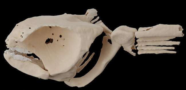
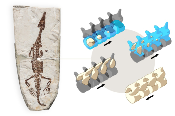
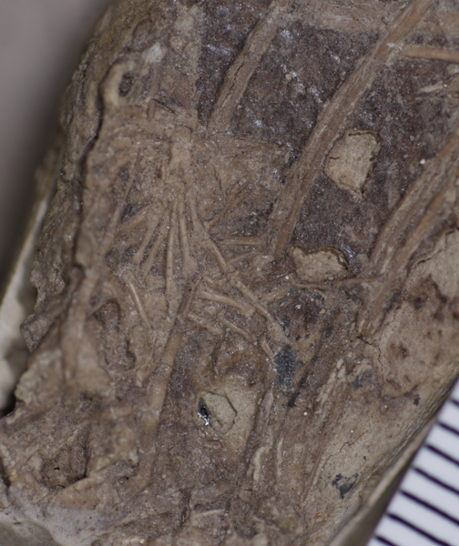
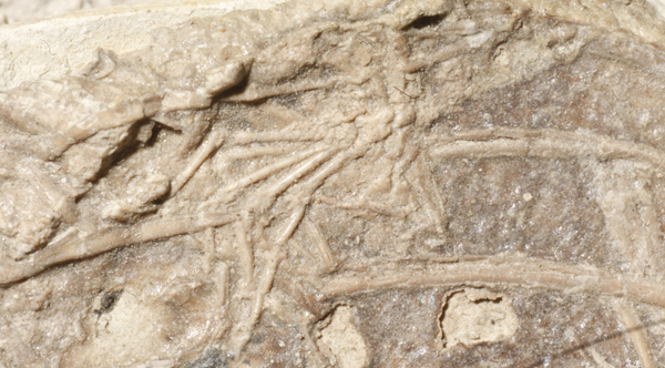
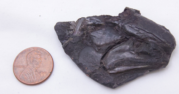
The Late Carboniferous (early Pennsylvanian subperiod), fish fossil was discovered in a layer of soapstone adjacent to a coal seam at the Mountain Fourfoot coal mine in Lancashire and the specimen was first scientifically described in 1925. The fish, named Coccocephalus wildi, would have measured around 20 cm in length and it lived in what was an ancient estuary. It is only known from this single fossil and only the skull and jaws were recovered.

Coccocephalus wildi fossil skull and jaws
The fossilised skull of Coccocephalus wildi. The fish is facing to the right, with the jaws visible in the lower right portion of the fossil. The eye socket is the circular, bumpy feature above the jaws. Picture credit: Jeremy Marble, University of Michigan News.

Vertebrate Brain Evolution

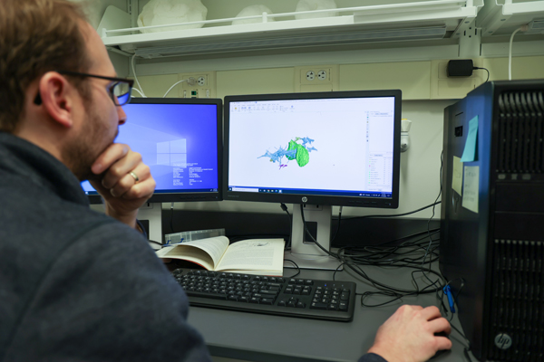
Coccocephalus was a member of the Class Actinopterygii, also known as the ray-finned fishes. The skull fossil was sent on loan from Manchester Museum to the University of Michigan and subsequent CT scans of the skull revealed the surprising discovery of the intact brain and associated nerves.

Senior author Sam Giles, (University of Birmingham), commented:

“This unexpected find of a three-dimensionally preserved vertebrate brain gives us a startling insight into the neural anatomy of ray-finned fish. It tells us a more complicated pattern of brain evolution than suggested by living species alone, allowing us to better define how and when present day bony fishes evolved.”

CT scans of C. wildi shedding light on vertebrate brain evolution.
University of Michigan palaeontologist Matt Friedman examines CT scan images of an exceptionally preserved, brain of the Late Carboniferous ray-finned fish Coccocephalus wildi. Picture credit: Jeremy Marble, University of Michigan News.

Rapidly Buried

When the fish died, it was probably buried rapidly in sediment containing very little oxygen. The lack of oxygen prevented the soft brain tissue from decaying. Whilst brain cases can reveal the shape and structure of vertebrate brains, this remarkable fossil preserved the brain tissue of a prehistoric fish.

Soft tissues such as the brain normally decay quickly and very rarely fossilise. But when this fish died, the soft tissues of its brain and cranial nerves were replaced during the fossilisation process with a dense mineral that preserved, in astonishing detail, their three-dimensional structure.

This discovery provides palaeontologists with a window into the evolution and development of the brains of ray-finned fishes, a highly successful group of back-boned animals estimated to represent more than fifty percent of all living vertebrate species.

Coccocephalus wildi life reconstruction and close-up view of brain. New study highlights vertebrate brain evolution.
Life reconstruction of the ray-finned fish Coccocephalus wildi showing location and shape of brain and cranial nerves. Picture credit: Márcio L. Castro.

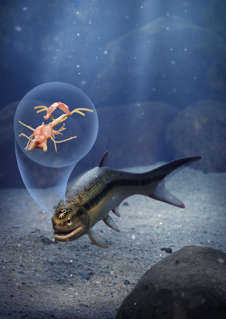
A study of the jaws and teeth of C. wildi suggest that it was carnivorous, likely feeding on small invertebrates. The CT scans revealed that the brain had bilateral symmetry, like the brains of modern ray-finned fishes, but significantly, the brain of Coccocephalus folds inward, unlike in all living ray-finned fishes, in which the brain folds outward.

For figures and replicas of ancient prehistoric fish: Prehistoric Sharks, Dunkleosteus and Other Prehistoric Fish Models (PNSO).

The fossil captures a time before a signature feature of ray-finned fish brains evolved, providing an indication of when this trait evolved.

Co-author of the paper, published in the journal “Nature”, Matt Friedman (University of Michigan) explained:

“An important conclusion is that these kinds of soft parts can be preserved, and they may be preserved in fossils that we’ve had for a long time—this is a fossil that’s been known for over 100 years.”

Everything Dinosaur acknowledges the assistance of a media release from the University of Birmingham in the compilation of this article.

The scientific paper: “Exceptional fossil preservation and evolution of the ray-finned fish brain” by Rodrigo T. Figueroa, Danielle Goodvin, Matthew A. Kolmann, Michael I. Coates, Abigail M. Caron, Matt Friedman and Sam Giles published in Nature.

30 01, 2023

A New Pterosaur Species is Described

By |2023-02-03T09:26:23+00:00January 30th, 2023|Categories: Dinosaur and Prehistoric Animal News Stories, Dinosaur Fans, Main Page, Palaeontological articles, Photos/Pictures of Fossils|0 Comments

A new pterosaur species has been described based on a superbly preserved specimen found in Upper Jurassic limestone deposits in Bavaria (southern Germany). The fully articulated specimen displays a unique dentition that suggests this flying reptile fed like a modern-day flamingo, sieving water through its jaws to trap small invertebrates as it waded or possibly swam in a shallow lagoon.

Balaenognathus Life Reconstruction
A life reconstruction of the newly described pterosaur Balaenognathus maeuseri. Picture credit: Megan Jacobs

Picture credit: Megan Jacobs

Balaenognathus maeuseri

The pterosaur has been classified as a ctenochasmatid, a group of short-tailed pterodactyloids characterised by specialised teeth adapted for filter feeding. Fossils of these relatively small flying reptiles (most with wingspans less than 3 metres), have been found in Europe, America and China, in rocks dating from the Upper Jurassic to the Early Cretaceous. The new pterosaur has been named Balaenognathus maeuseri, the genus name derives from the scientific name for the Bowhead whale (Balaena mysticetus) and the Latin for jaw, as it is thought that these two unrelated species shared a common feeding strategy. The specific epithet honours a co-author of the paper Matthias Mäuser who sadly passed away before publication.

Balaenognathus fossil specimen.
The fossilised bones of Balaenognathus maeuseri found in the slab of limestone (Upper Jurassic laminated limestones at Wattendorf, Bavaria in Southern Germany). Picture credit: PalZ.

Lead author of the study, published in Paläontologische Zeitschrift (PalZ), Professor David Martill from the University of Portsmouth School of the Environment, Geography and Geosciences commented:

“The nearly complete skeleton was found in a very finely layered limestone that preserves fossils beautifully.”

Unique Pterosaur Dentition

The fossil (specimen number NKMB P2011-63), is remarkable for its completeness, unusual dentition and hints of the preservation of soft tissues, including wing membranes. The delicate jaws contain at least 480 fine teeth.”

Professor Martill added:

“The jaws of this pterosaur are really long and lined with small fine, hooked teeth, with tiny spaces between them like a nit comb. The long jaw is curved upwards like an avocet and at the end it flares out like a spoonbill. There are no teeth at the end of its mouth, but there are teeth all the way along both jaws right to the back of its smile.”

Tentative line reconstruction of the skull.
Tentative line reconstruction of the skull. Picture credit: PalZ

Bizarre Hook-like Tooth Crown

The tips of the jaw are devoid of teeth, which would have permitted plankton and invertebrate-rich water to rush into the long jaw. The hundreds of teeth would have acted as a sieve helping to strain out food. Many of the teeth have a hook-like expansion on the tip of the crown, a bizarre and unique tooth morphology.

Explaining the significance of these strange teeth, Professor Martill stated:

“What’s even more remarkable is some of the teeth have a hook on the end, which we’ve never seen before in a pterosaur ever. These small hooks would have been used to catch the tiny shrimp the pterosaur likely fed on – making sure they went down its throat and weren’t squeezed between the teeth.”

Balaenognathus teeth with hook-like crowns.
Fig 7 shows UV images of the teeth (A) teeth close to the tip of the jaw (B) close-up of the crown tips of the teeth of the left jaw showing the hook-like teeth with the hooks highlighted by white arrows. Image (C) the middle teeth. Picture credit: PalZ.

A New Pterosaur

The discovery was made accidentally while scientists were excavating a large block of limestone containing crocodilian fossil remains.

Professor Martill explained:

“This was a rather serendipitous find of a well-preserved skeleton with near perfect articulation, which suggests the carcass must have been at a very early stage of decay with all joints, including their ligaments, still viable. It must have been buried in sediment almost as soon as it had died.”

Most members of the Ctenochasmatidae family seem to have been the pterosaur equivalent of wading shore birds, although some genera were perhaps adapted to habitats further inland and have truly bizarre shaped jaws leaving palaeontologists perplexed as to what they ate.

Only one other known pterosaur had more teeth than Balaenognathus. It is another ctenochasmatid and it is called Pterodaustro guinazui and its fossils are known from the Lower Cretaceous of Argentina. Both Pterodaustro and Balaenognathus were likely filter feeders although the arrangement of their teeth differs. Balaenognathus had teeth in the upper and lower jaw which are the mirror image of each other, whilst P. guinazui had very reduced teeth in the upper jaw and up to a 1,000 densely packed, bristle-like teeth in the lower jaw.

Balaenognathus fossil specimen viewed under UV light.
The Balaenognathus maeuseri specimen viewed under UV (ultra violet) light. Picture credit: PalZ.

New Pterosaur Species – Unique Feeding Mechanism

The teeth of Balaenognathus suggest a feeding strategy that involved the animal either wading through water or swimming, using its spoon-shaped beak to funnel water into its mouth, this water was then strained through its teeth to trap prey. The researchers propose that Balaenognathus fed on shrimps and copepods filling a similar ecological niche as extant ducks, shorebirds and flamingos.

Commenting on the sad passing of Matthias Mäuser, Professor Martill said:

“Matthias was a friendly and warm-hearted colleague of a kind that can be scarcely found. In order to preserve his memory, we named the pterosaur in his honour.”

Everything Dinosaur acknowledges the assistance of a media release from the University of Portsmouth in the compilation of this article.

The scientific paper:

The scientific paper: “A new pterodactyloid pterosaur with a unique filter‑feeding apparatus
from the Late Jurassic of Germany”
by David M. Martill, Eberhard Frey, Helmut Tischlinger, Matthias Mäuser, Héctor E. Rivera‑Sylva and Steven U. Vidovic published in Paläontologische Zeitschrift (PalZ).

16 01, 2023

A New Study – Carboniferous Chimaeras were Suction Feeders

By |2024-01-02T13:59:46+00:00January 16th, 2023|Categories: Adobe CS5, Dinosaur and Prehistoric Animal News Stories, Dinosaur Fans, Main Page, Palaeontological articles, Photos/Pictures of Fossils|0 Comments

Carboniferous chimaeras were suction feeders unlike their modern relatives such as the rat fish which are durophagous (feed on hard-shelled prey such as crabs, snails and molluscs). That is the conclusion of new research published this week in the academic journal The Proceedings of the National Academy of Sciences (PNAS).

An Exceptional Three-dimensional Fossil

The research led by the Muséum national d’histoire naturelle (MNHN) located in Paris, and the University of Birmingham has shown that an ancient relative of chimaeras, jawed vertebrates that are related to cartilaginous fishes (sharks and rays), fed by sucking in prey animals underwater.

An exceptional three-dimensional fossil of an ancient chimaera (Iniopera genus), has revealed new clues about the diversity of these creatures during the Carboniferous period.

Carboniferous chimaeras
The three-dimensional cast of the Carboniferous chimaera fossil (Iniopera) which helped the researchers to determine feeding strategy. Picture credit: University of Birmingham.

Carboniferous Chimaera

The fossil, from a genus called Iniopera, is the only suction feeder to be identified among chimaeras, and quite different from living chimaeras, which generally feed by crushing molluscs and other hard-shelled prey between their teeth.

Chimaeriformes are an ancient order of cartilaginous fish (Chondrichthyes) that are thought to have evolved in the Devonian. Most extant species are found at depths greater than two hundred metres, and some chimaera fish are restricted to extremely deep water (Bathypelagic Zone).

Most fossil and extant chimaeras are quite small, very few specimens exceed one metre in length. However, other prehistoric, cartilaginous fish that were distantly related to Iniopera grew much larger. For example, the Permian genus Helicoprion with its bizarre tooth-whorl jaw, which has been estimated to have grown to around eight metres in length.

Helicoprion scale drawing
As Everything Dinosaur prepares for the arrival of Haylee the Helicoprion model from PNSO a scale drawing of this Permian fish has been commissioned. Picture credit: Everything Dinosaur.

Picture credit: Everything Dinosaur

Although models of prehistoric fish from the Chondrichthyes Class are rare, PNSO have included two prehistoric shark figures (O. megalodon and Cretoxyrhina) and a replica of Helicoprion.

To view the PNSO prehistoric animal model range in stock at Everything Dinosaur: PNSO Age of Dinosaurs Models and Figures.

Identifying a Suction Feeder

Commenting on the significance of this study, lead researcher Dr Richard Dearden (University of Birmingham) stated:

“Being able to identify Iniopera as a suction feeder sheds light on the diverse role of chimaeras in these early ecosystems. In particular, it suggests that in their early evolutionary history, some chimaeras were inhabiting ecological niches that are now monopolised by ray finned fishes – a far cry from their modern life as specialised shell-crushers.”

The cartilage skeleton of these fish are rarely fossilised and the Chondrichthyes tend to be underrepresented in the fossil record. The skeletons that are preserved tend to be crushed flat and distorted so interpreting them is notoriously difficult. However, by studying the tooth shapes and diverse body plans, palaeontologists were already aware that extinct forms were far more varied than their living counterparts.

3-D Imaging Techniques

Using advanced 3-D imaging techniques, the researchers reconstructed the head, shoulder and throat skeleton of the Iniopera fossil. They then estimated the location of major muscles and found the anatomy was poorly suited to durophagous feeding. Instead, the researchers believe the animal was more likely to have used the muscle arrangement to expand the throat to take in water and a forward-pointing mouth to orient towards prey.

Suction feeding is a technique used by many animals that live underwater. It involves generating low pressures in the throat to pull in water and prey. To do this effectively, the animal needs to be able to rapidly expand its throat, and point its mouth forward towards prey items. Numerous different aquatic jawed vertebrates, such as ray-finned fishes and some turtles have evolved specialised anatomies to help them feed in this manner more effectively.

The suction feeding theory is also supported by fossilised Chimaeriformes that have preserved stomach contents. Small arthropods have been found in association with the body cavity of several specimens and their relatively entire state suggests suction feeding as the method of prey capture.

Everything Dinosaur acknowledges the assistance of a media release from the University of Birmingham in the compilation of this article.

5 01, 2023

The Amazing Skin of Diplodocus

By |2024-01-02T14:02:44+00:00January 5th, 2023|Categories: Adobe CS5, Dinosaur and Prehistoric Animal News Stories, Dinosaur Fans, Main Page, Palaeontological articles|0 Comments

The debate over whether theropod dinosaurs were feathered, scaly or perhaps a combination of both continues. In contrast, the integumentary covering of sauropod dinosaurs has largely remained uncertain due to the very limited fossil evidence. However, at one remarkable location in Montana, patches of fossilised Diplodocus skin have been preserved and an analysis indicates that these long-necked dinosaurs were most likely covered in scales, but surprisingly their skin scales were not uniform. A range of scale shapes are indicated, all of them relatively small, but ovoid, rectangular, polygonal, domed scales and irregular (globular) scales have been identified.

The texture on the neck and shoulders of the Schleich Brachiosaurus dinosaur model.
The scales found on a replica of the sauropod Brachiosaurus (Schleich) research suggests that diplodocids were covered in a variety of scales. Globular and domed scales have been identified along with ovoid and more uniform polygonal scales. Picture credit: Everything Dinosaur.

A Research Paper

In a scientific paper published in the on-line, open-access journal PeerJ (April 2021), written by Tess Gallagher (currently a Masters student at Bristol University), in collaboration with colleagues from, what was known at the time as the Bighorn Basin Paleontological Institute, descriptions were provided of some patches of skin associated with juvenile Diplodocus fossils.

The fossil material comes from a single location known as the Mother’s Day Quarry, located in the Bighorn Basin, (Montana). The site was originally discovered in the 1990s and the Upper Jurassic deposits have yielded over two thousand Diplodocus fossil bones. Surprisingly, given the concentration of sauropod material found, very few other fossils have been discovered in this quarry. To date, some theropod teeth (allosaurid) are known plus one potential theropod footprint, preserved in association with the skin and a single invertebrate specimen.

It has been suggested that a herd of young Diplodocus dinosaurs died from lack of water at a dried up watering hole. Their bodies remained on the surface for some time and became desiccated. A flash flood occurred and swept the corpses downstream and these accumulated bodies were rapidly buried.

The dinosaur skin, having been bleached and dried out on the surface, once rapidly buried has an increased chance of becoming fossilised. Recently published research (October 2022), from Drumheller et al suggests that “mummified” skin might be more common in the fossil record than previously thought.

To read Everything Dinosaur’s blog post about this phenomenon: Dinosaur Mummies an Alternative Fossil Pathway.

Fossilised Diplodocus Skin

Although only a small portion of skin has been identified to date and the scientists remain uncertain as to which part of the body the skin patches covered, analysis has revealed a remarkably diverse quantity of scales. The team conclude that considering how diverse the scale shapes are in such a small area of skin, it is possible that these distinct scale shapes may represent a transition on the body from one region to another, perhaps from the abdomen to the dorsal side, or abdomen to the shoulder.

Fossilised Diplodocus skin.
Photograph of specimen number MDS-2019-028 showing patches of diplodocid skin preserved between two rib bones. The black arrow indicates north. Picture credit: Gallagher, Poole and Schein.

At least six different types of Diplodocus scale have been identified, suggesting that the skin of these sauropod dinosaurs was complex. In addition, a further study presented at the recent annual meeting of the Society of Vertebrate Paleontology in Canada, provided a fresh perspective on the specialised skin of these sauropods.

Analysis of the scales using a high-powered microscope revealed that they had unusually large pores. It has been proposed that these large pores helped increase the surface area of this huge dinosaur. The greater the surface area of the skin the more assistance it would have been in helping to keep Diplodocus cool. Elephants have wrinkled skin, this increases the skin’s surface area which helps to minimise the impact of the “square-cube law” in relation to overheating.

“Square-cube” Law

The larger the volume of an animal, the smaller the overall surface area of the skin in contact with the air and this makes dissipating heat difficult for large animals. Just as elephants help to resolve issues with overheating with their wrinkled skin, the large pores associated with the Diplodocus skin had a similar effect.

Fossilised Diplodocus skin (line drawing).
Simplified line drawing showing the distribution of different scale shapes on the specimen number MDS-2019-028. The black arrow shows north. Drawing by T. Gallagher. Picture credit: Gallagher, Poole and Schein.

The scientific paper: “Evidence of integumentary scale diversity in the late Jurassic Sauropod Diplodocus sp. from the Mother’s Day Quarry, Montana” by Tess Gallagher, Jason Poole and Jason P. Schein published in PeerJ.

4 01, 2023

New Study Shows Plant-eating Dinosaurs Ate Plants Differently

By |2024-01-02T14:02:28+00:00January 4th, 2023|Categories: Dinosaur and Prehistoric Animal News Stories, Main Page, Palaeontological articles|0 Comments

Newly published research demonstrates that plant-eating, ornithischian dinosaurs had different ways of tackling the plants that made up their diet. Scans of the skulls of five, herbivorous dinosaurs, all members of the bird-hipped group (Ornithischia), were used to create three-dimensional models of the skull, teeth and jaws. These computer models were then subjected to a series of stress tests measuring the jaw muscles and calculating bite forces to help palaeontologists understand how different feeding strategies evolved in the Dinosauria.

Life-size Psittacosaurus replica.
A model of the dinosaur called Psittacosaurus. A skull model of this Early Cretaceous dinosaur was tested to determine the impact of bite force stresses on the bones. This data provided the scientists with information on different feeding strategies within the Ornithischia. Picture credit: Everything Dinosaur.

Picture credit: Everything Dinosaur

Plant-eating Dinosaurs

It is thought that the very earliest dinosaurs were carnivorous. However, quite early in their evolutionary history, the Dinosauria diversified and new forms with different diets (herbivory and omnivory) evolved.

In a recently published study (December 2022), analysis of dinosaur tooth shape suggested that the ancestors of the huge, herbivorous sauropods were meat-eaters, whilst many groups of plant-eating, ornithischian dinosaurs were ancestrally omnivorous.

To read Everything Dinosaur’s blog post about this research: Tooth Shape Helps Shape Dinosaur Diet.

Earliest Representatives of Major Ornithischian Groups

The skull and jaw muscles of some of the earliest representatives of major families within the Ornithischia were studied namely:

  • Heterodontosaurus – Heterodontosauridae family from the Early Jurassic.
  • Lesothosaurus – A basal ornithischian known from the Early Jurassic, possibly part of the early ornithopod lineage or perhaps an ancestor of armoured dinosaurs (Thyreophora).
  • Scelidosaurus – An early member of the Thyreophora (Early Jurassic).
  • Hypsilophodon – Regarded as a basal ornithopod (Early Cretaceous).
  • Psittacosaurus – A basal member of the Marginocephalia clade (Early Cretaceous) which includes horned dinosaurs (ceratopsids) and the bone-headed dinosaurs (pachycephalosaurs).

Writing in the academic journal “Current Biology”, the research team, which included scientists from the University of Birmingham, the London Natural History Museum and Bristol University, conclude that these herbivorous dinosaurs evolved very different ways of tackling their diet of vegetation.

Plant-eating dinosaurs ate plants differently.
Different feeding strategies in ornithischian dinosaurs. Computerised tomography was used to create models of skulls and these models were subjected to bite force stress tests to assess how these dinosaurs fed. Picture credit: David Button.

Skull Morphology and Jaw Musculature Reveal Different Feeding Strategies

Using computer models and finite element analysis to assess the impact of stress on the skull and bite forces the team discovered that Heterodontosaurus had disproportionately large jaw muscles in relation to the size of its skull. It had a powerful bite. As it was able to generate a higher bite force this would have helped it to consume tough plants. Scelidosaurus had a similar bite force, but relatively smaller jaw muscles compared to the size of its skull. Hypsilophodon, in contrast, had proportionately smaller jaw muscles, it could bite more efficiently but with less force.

Co-author of the study, Dr Stephan Lautenschlager (University of Birmingham), commented:

“We discovered that each dinosaur tackled the problems posed by a plant-based diet by adopting very different eating techniques. Some compensated for low eating performance through their sheer size, whilst others developed bigger jaw muscles, increased jaw system efficiency, or combined these approaches. Although these animals looked very similar, their individual solutions to the same problems illustrates the unpredictable nature of evolution.”

Compared to Birds and Crocodilians

The jaw muscles were reconstructed on the model skulls using extant archosaurs as templates (birds and crocodilians). Finite element analysis was then conducted to determine the potential bite force of each dinosaur. Finite element analysis involved dividing the skull into thousands of individual parts (called elements). The bite force these muscles can generate is calculated based on their size and arrangement.

Heat maps showed the different stress levels generated throughout each skull as the biting motion was simulated. The results revealed that although all of these dinosaurs were eating plants, each type of dinosaur had a different way of doing it.

Professor Paul Barrett (London Natural History Museum), explained that it was essential for palaeontologists to understand how dinosaurs evolved to feed on plants in so many ways. This diversity in feeding strategies helps to explain how these animals came to be the dominant primary consumers in terrestrial food chains for millions of years.

Lead author of the study, Dr David Button (University of Bristol) explained:

“When we compared the functional performance of the skull and teeth of these plant-eating dinosaurs, we found significant differences in the relative sizes of the jaw muscles, bite forces and jaw strength between them. This showed that these dinosaurs, although looking somewhat similar, had evolved very different ways to tackle a diet of plants.”

Scelidosaurus
An illustration of the Early Jurassic armoured dinosaur Scelidosaurus. A study of this dinosaur’s skull morphology and jaw muscles has led to palaeontologists gaining a new perspective on the feeding strategies of early armoured dinosaurs. Picture credit: Everything Dinosaur.

Dr Button went onto add:

“This research helps us understand how animals evolve to occupy new ecological niches. It shows that even similar animals adopting similar diets won’t always evolve the same characteristics. This highlights how innovative and unpredictable evolution can be.”

These differences in feeding strategy identified in this research demonstrates that each of these types of ornithischian dinosaur evolved a distinct solution to feeding on plants.

Everything Dinosaur acknowledges the assistance of a media release from the University of Birmingham in the compilation of this article.

The scientific paper: “Multiple pathways to herbivory underpinned deep divergences in ornithischian evolution” by David J. Button, Laura B. Porro, Stephan Lautenschlager, Marc E. H. Jones and Paul M. Barrett published in Current Biology.

2 01, 2023

Sauropod Dinosaurs Did Not Have Supersonic Tails

By |2023-01-03T07:45:19+00:00January 2nd, 2023|Categories: Adobe CS5, Dinosaur and Prehistoric Animal News Stories, Dinosaur Fans, Main Page, Palaeontological articles|0 Comments

A recent study published in the academic journal “Scientific Reports” refutes the idea that some long-necked herbivores had supersonic sauropod tails. The controversial idea that some dinosaurs could lash their tails like a whip creating a supersonic crack as the tail travelled faster than the speed of sound has been refuted in newly published research. Instead, the researchers suggest that the tail of diplodocids such as Apatosaurus, Brontosaurus and Diplodocus could still play a role in defence, producing a painful blow to deter an attacker. It is also suggested that these long, whip-like tails could have been used in intraspecific combat.

Apatosaurus scale drawing.
Scale drawing of Apatosaurus (A. ajax). Note the long, whip-like tail. New research suggests that these long tails could not be used to create a “crack” as they broke the sound barrier. Picture credit: Everything Dinosaur.

Picture credit: Everything Dinosaur

Supersonic Sauropod Tails

A sauropod clade, the Flagellicaudata are characterised by their extremely long tails. This clade includes the Diplodocidae family and the closely related Dicraeosauridae. Although complete fossil sauropod tails are extremely rare, palaeontologists have a good idea of the anatomy of a typical diplodocid tail. It consisted of approximately eighty caudal vertebrae, that gradually decrease in size and morphological complexity towards the tail tip. There are approximately ten larger posterior vertebrae, followed by forty or so intermediate bones with finally around thirty progressively smaller rod-like caudal vertebrae.

Earlier studies had suggested that the tail could be whipped, and the tip would travel so fast (in excess of 500 metres per second), this action would break the sound barrier and produce a loud sound. This speedy tail would cause a significant injury should it come into contact with another dinosaur.

However, this new study used three-dimensional models and computer analysis to assess the stress on the bones, ligaments and soft tissues. They concluded that the maximum tip velocity generated would be around thirty metres a second, nowhere near the 330 metres per second required to break the sound barrier.

Eofauna Diplodocus scale model
The Eofauna Diplodocus carnegii model measures around 60 cm in length and stands 11 cm tall. It is a 1/40th scale model. Most of the model’s length is made up of the long tail. Diplodocids are members of the Flagellicaudata clade.

The picture (above) shows the recently introduced Eofauna Scientific Research Diplodocus carnegii replica. When shown in lateral view, the extremely long tail can be seen.

To view the range of models and figures in the Eofauna series: Eofauna Scientific Research Models.

An Effective Weapon

Whilst the researchers conclude that the effect of friction on the musculature and aerodynamic drag would prevent the tail tip from reaching a speed capable of breaking the sound barrier, the pressure applied by the terminal section would not be enough to break bones or lacerate dinosaur skin, but it could still deliver a painful blow.

In summary, the scientists suggest that sauropod tail use remains speculative, these tails could have been used in intraspecific combat, or perhaps as a weapon against predators. Similarly, the use of the tail as a tactile element to retain herd cohesion is equally plausible.

The scientific paper: “Multibody analysis and soft tissue strength refute supersonic dinosaur tail” by Simone Conti, Emanuel Tschopp, Octávio Mateus, Andrea Zanoni, Pierangelo Masarati and Giuseppe Sala published in Scientific Reports.

2 01, 2023

The Evolution of the Backbone

By |2022-12-30T14:49:20+00:00January 2nd, 2023|Categories: Adobe CS5, Dinosaur and Prehistoric Animal News Stories, Main Page, Palaeontological articles|0 Comments

The evolutionary development of the vertebral column has been extensively researched. Numerous fossil specimens have been studied as scientists pursue a greater understanding of the evolution of the backbone. Recently, a new scientific paper has been published in “Scientific Reports” that outlines the evolutionary development of ossification patterns in four-legged vertebrates.

Research from the Museum für Naturkunde

The study was undertaken by scientists from the Museum für Naturkunde (Berlin, Germany). Antoine Verrière and his colleagues were able to reconstruct the patterns of how the bones in the vertebral column formed in the ancestor to all land vertebrates based on a large dataset compiled from studies of extant and extinct vertebrates. The dataset also included new information on the spine of Mesosaurus tenuidens, widely regarded as the first reptile to adapt to an aquatic existence, back in the Permian some 300 million years ago.

Evolution of the Backbone.
Understanding the evolution of ossification patterns in the backbones of four-legged vertebrates. Picture credit: Verrière and Fröbisch.

The Evolution of the Backbone

Lead author of the paper, Antoine Verrière explained that M. tenuidens had a long snout and a powerful tail that propelled it through the water. It inhabited an inland sea that once existed in the southern region of the supercontinent Pangaea.

The palaeontologist added:

“On some rare juvenile specimens, we observed that the neural arches, the spines sitting on top of the main part of a vertebra, were closing from head to tail as the animals grew, much like a zipper. We wanted to understand how this pattern would fit in the evolutionary history of land vertebrates, but quickly realised there was surprisingly little information available. So, we decided to investigate this ourselves!”

Four Major Developmental Patterns in Backbones of Amniotes

The research team looked at four of the major developmental patterns in the backbones of amniotes (mammals, reptiles and birds):

  • The ossification of the centrum (the main body of a vertebra).
  • The ossification of paired neural arches.
  • The fusion of the initially forming paired neural arch elements into one spine.
  • The fusion of neural arches with the centrum, also called neurocentral fusion.

Statistical analysis was used to model how these different patterns changed from the Permian through to today, their work roughly covering the evolutionary history of land-living vertebrates excluding amphibians. With this research the team could reconstruct the patterns in the common ancestor to all land vertebrates.

Co-author of the study, Professor Jörg Fröbisch (Museum für Naturkunde) commented:

“What surprised us the most was that these patterns appear to have been relatively stable for the last 300 million years. Modern and extinct vertebrates are enormously diverse in terms of their body shapes and lifestyles and the elements of their vertebral columns are organised in complex units that differ greatly between species. Nevertheless, the ossification patterns were much more conservative than was expected from the great morphological diversity.”

Edmontosaurus skeleton.
Duck-billed dinosaur on display showing the vertebral column. Despite vertebrates having extremely diverse body shapes and complex spines the observed ossification patterns were much more conservative than expected. Picture credit: Everything Dinosaur.

Picture credit: Everything Dinosaur

Some Deviations Identified

Although the patterns studied show relative stability through deep geological time, some deviations were identified. Notably, birds, mammals, and members of the Squamata Order (snakes and lizards) each evolved their own specific modes of vertebral ossification, which differ from the ancestral condition in amniotes. Yet again, within these groups, the patterns were also surprisingly stable.

Fellow co-author Professor Nadia Fröbisch (Museum für Naturkunde) explained:

“Ostriches and seagulls, for instance, have very different anatomies and lifestyles, but their vertebral columns ossify in similar ways. This shows that some changes can be observed between the major lineages of land vertebrates, but within each of the main lineages, spine development remained rather stable again.”

This study demonstrates how studying modern animals alongside their ancient ancestors can provide a much deeper understanding of the evolutionary development of key anatomical structures.

Everything Dinosaur acknowledges the assistance of a media release from the Museum für Naturkunde (Berlin) in the compilation of this article.

The scientific paper: “Regionalization, constraints, and the ancestral ossification patterns in the vertebral column of amniotes” by Antoine Verrière, Nadia B. Fröbisch and Jörg Fröbisch published in Scientific Reports.

29 12, 2022

A New Dwarf Nodosaurid Called Patagopelta

By |2024-02-08T08:38:09+00:00December 29th, 2022|Categories: Dinosaur and Prehistoric Animal News Stories, Dinosaur Fans, Main Page, Palaeontological articles, Photos/Pictures of Fossils|0 Comments

In today’s blog post we look at the dwarf nodosaurid Patagopelta (P. cristata), which was formally named and described earlier this month.

A new, very small, armoured dinosaur has been named and described from fossils found in Argentina. The dinosaur which measured around 2 to 2.3 metres in length (based on the dimensions of the femur), suggests that some members of the Nodosauridae in Gondwana became smaller in the Late Cretaceous, perhaps as armoured dinosaurs in South America were under evolutionary pressure from other ornithischians and titanosaurs.

Dwarf nodosaurid Patagopelta
A life reconstruction of the newly described, dwarf nodosaurid from Argentina (Patagopelta cristata). Picture credit: Gabriel Diaz Yantén.

Dwarf Nodosaurid Patagopelta

Fragmentary remains of Late Cretaceous armoured dinosaurs are known from Chile and Argentina, but little work had been undertaken to assess these specimens and to review their phylogeny and taxonomic relationship with other members of the Ankylosauria clade from North America and elsewhere in the world.

Writing in the ” Journal of Systematic Palaeontology”, the researchers led by Facundo Riguetti, a CONICET doctoral fellow, reassessed the known ankylosaur material in conjunction with some other recently found fossils and, as a result, they were able to establish a new nodosaurid species from bones and a single tooth found in sediments of the Allen Formation (Campanian–Maastrichtian) in Salitral Moreno, Río Negro Province (northern Patagonia).

Patagopelta cristata

The dinosaur’s genus name translates as “Patagonian shield” whilst the trivial name derives from the Latin for crest – a reference to the diagnostic crests on both the anterior surface of the femur and the lateral osteoderms of the cervical rings.

Dr Riguetti commented:

“The importance of the study lies in the fact that Patagopelta is the first species of Ankylosauria described for the continental territory of Argentina, which fills the existing gap for this group and adds a new thyreophoran to the very few incomplete and indeterminate remains known for our country from this type of ornithischian dinosaur.”

Dwarf nodosaurid Patagopelta (views of the femur).
The right femur of Patagopelta (specimen number MPCA-SM-1), in A, anterior, B, posterior, C, lateral, D, medial, E, proximal and F, distal views. As the fragmentary left femur would have been the same size it is thought the femora came from a single animal. Other fossil remains represent several individuals. Abbreviations: fh, femoral head; fn, fibular notch; ft, fourth trochanter; gtr, greater trochanter; it, interwoven texture; lc, lateral condyle; le, lateral epicondyle; li (atr), linea intermuscular (associated to the anterior trochanter); lmca, linea muscularis caudalis; lmcr, linea muscularis cranialis; mc, medial condyle. Scale bar = 10 cm. Picture credit: Riguetti et al.

The Right Femur

The best-preserved fossil element is the right femur, which is complete and shows typical anatomical characteristics associated with the Nodosauridae. This bone along with the distinctive cervical osteoderms led to the erection of this new species. As the femur is only 25 cm in length and bone histology suggests an adult animal, the researchers conclude that Patagopelta was a dwarf form of armoured dinosaur.

Co-author Sebastián Apesteguía, a CONICET researcher, explained:

“For an armoured dinosaur, Patagopelta is extremely small. Due to the size of the femur, only 25 centimetres in length, we estimate that the animal must have been between two and three meters long, while, in general, ankylosaurs are medium-sized or large animals, with an average length of between four and five metres.”

A Faunal Exchange Across the Americas

Although it is thought that the Nodosauridae evolved in the Northern Hemisphere, towards the end of the Cretaceous (Campanian – Maastrichtian), a land bridge existed between North America and South America that permitted a faunal exchange. Titanosaurs migrated north, which explains why fossils of titanosaurs such as Alamosaurus occur in the USA. Ornithischian dinosaurs such as hadrosaurs and nodosaurids moved south.

Alamosaurus scale drawing.
Scale drawing of Alamosaurus. A giant titanosaur known from North America that is probably descended from titanosaurs that roamed South America. Picture credit: Everything Dinosaur.

The image above shows a typical Late Cretaceous titanosaur, for models of Late Cretaceous dinosaurs including titanosaurs and armoured dinosaurs: CollectA Prehistoric Life Models.

Sebastián Apesteguía added:

“That is why in South America we only expect to find animals like Patagopelta in rocks from the Late Cretaceous, just before the global extinction of the dinosaurs took place.”

Dwarfism in Late Cretaceous South American Thyreophora

The size of Patagopelta along with the recently described Stegouros (Soto-Acuña et al, 2021)*, from southernmost Chile, suggests that armoured dinosaurs in South America may have gradually become smaller. This trait is not known in members of the Thyreophora described from other parts of the world. Palaeontologists have speculated that perhaps competition from titanosaurs and the migration of hadrosaurs into South America might have led to armoured dinosaurs adapting to different ecological niches to avoid competition. By being smaller these animals needed fewer resources than larger, contemporaneous herbivorous dinosaurs.

It has also been suggested that the geology of Patagonia where the fossils of Patagopelta were found might provide a clue to the dwarfism. Geologists are aware of several Late Cretaceous marine transgressions in the region. This might have led to the establishment of an island archipelago with dinosaurs living on these small islands gradually become smaller due to a scarcity of resources (the “island rule”).

Tracks of Dwarf Ankylosaurs

Members of the Patagopelta research team had previously described tracks of dwarf ankylosaurs, possibly affected by similar circumstances, preserved in Upper Cretaceous deposits in Bolivia.

*To read Everything Dinosaur’s 2021 article about the discovery of Stegouros: New Armoured Dinosaur from Chile.

Everything Dinosaur acknowledges the assistance of a media release from Consejo Nacional de Investigaciones Científicas y Técnicas (CONICET) in the compilation of this article.

The scientific paper: “A new small-bodied ankylosaurian dinosaur from the Upper Cretaceous of North Patagonia (Río Negro Province, Argentina)” by Facundo Riguetti, Xabier Pereda-Suberbiola, Denis Ponce, Leonardo Salgado, Sebastián Apesteguía, Sebastián Rozadilla and Victoria Arbour published in the Journal of Systematic Palaeontology.

23 12, 2022

New Study Suggests Tooth Shape Helps Shape Dinosaur Diet

By |2024-02-08T08:39:48+00:00December 23rd, 2022|Categories: Adobe CS5, Dinosaur and Prehistoric Animal News Stories, Dinosaur Fans, Main Page, Palaeontological articles|0 Comments

Recently published research examining tooth shape in early members of the Dinosauria has provided new information on dinosaur diet. The very earliest known dinosaurs included carnivores, herbivores as well as omnivores. Early dinosaurs were already developing adaptations to exploit a wide variety of feeding strategies.

Early dinosaur diet investigated.
Buriolestes (top left), a member of the Sauropodomorpha is a carnivore whilst the geologically younger sauropodomorph Thecodontosaurus is thought to have been herbivorous (top right). Lesothosaurus (bottom), an early member of the Ornithischia, is thought to be an omnivore. Picture credit: Gabriel Ugueto.

Picture credit: Gabriel Ugueto

Dinosaur Diet

Writing in the academic journal “Science Advances”, the scientists from the University of Bristol developed computer models to test the function and bite force of the teeth of a variety of early dinosaurs. These results were then compared with the data from extant lizards so that the diet could be inferred. The study shows that many groups of plant-eating ornithischian dinosaurs were ancestrally omnivorous and the ancestors of the huge sauropods, dinosaurs such as Apatosaurus, Diplodocus, Dreadnoughtus and Argentinosaurus were carnivores.

The scientists conclude that the ability of the Dinosauria to diversify their diets early in their evolution probably explains their evolutionary and ecological success.

Studying the Earliest Dinosaurs

The Dinosauria dominated terrestrial ecosystems for much of the Mesozoic. However, their origins and how they came to out compete other tetrapods during the Middle to Late Triassic remains the subject of intense debate. Over a few million years, the dinosaurs seem to have rapidly diversified and moved from being essential “bit-part” players in terrestrial ecosystems dominated by other types of archosaur and synapsid to becoming the dominant group.

Analysis of trackways discovered in the Southern Alps suggests a link between extensive faunal turnover leading to the dominance of the Dinosauria and the Carnian Pluvial Episode (CPE), a period of major climate change and a shift in the types of flora.

The diversification of the dinosaurs.
The diversification of the dinosaurs coincides with the Carnian Pluvial Episode (CPE). Picture credit: Everything Dinosaur.

Picture credit: Everything Dinosaur

To read more about this research into the impact of the Carnian Pluvial Episode on terrestrial ecosystems: Dinosaurs – In with a Bang and Out with a Bang.

A Wide Diversity of Different Skull and Tooth Shapes

Commenting on the implications of this study, lead author Dr Antonio Ballell stated:

“Soon after their origin, dinosaurs start to show an interesting diversity of skull and tooth shapes. For decades, this has made palaeontologists suspect that different species were already experimenting with different kinds of diets. They have compared them to modern lizard species and tried to infer what they ate based on the similarities in their teeth.”

Tooth morphology yields data on dinosaur diet.
Dinosaur skull shape and tooth morphology mapped over time. The three main dinosaur lineages, Sauropodomorpha, Ornithischia and Saurischia are represented from the Late Triassic to the Early Jurassic. Sauropodomorphs that were ancestral to the giant plant-eaters such as Apatosaurus and Dreadnoughtus were originally carnivorous, whilst ornithischian dinosaurs regarded as predominately herbivorous, started off as omnivores. Picture credit: Ballell, Benton and Rayfield.

Dr Ballell, based at the University’s School of Earth Sciences added:

“We investigated this by applying a set of computational methods to quantify the shape and function of the teeth of early dinosaurs and compare them to living reptiles that have different diets. This included mathematically modelling their tooth shapes and simulating their mechanical responses to biting forces with engineering software.”

A Plateosaurus dinosaur model.
A rearing Plateosaurus. The study confirmed that the large, Late Triassic Plateosaurus was in all probability a herbivore.

The Plateosaurus replica (above), is part of the CollectA not-to-scale range of prehistoric animal models.

To view this range: CollectA Prehistoric Life Models and Figures.

Predicting Dinosaur Diet

Co-author of the paper, Professor Mike Benton explained:

“With this battery of methods, we were able to numerically quantify how similar early dinosaurs were to modern animals, providing solid evidence for our inferences of diets. Theropod dinosaurs have pointy, curved and blade-like teeth with tiny serrations, which behaved like those of modern monitor lizards. In contrast, the denticulated teeth of ornithischians and sauropodomorphs are more similar to modern omnivores and herbivores, like iguanas.”

Innovative Machine Learning

This innovative research used machine learning models to group the earliest dinosaurs into different diet categories based on their jaw mechanics and tooth shape. For example, Thecodontosaurus, a dinosaur which roamed the Triassic archipelago where Bristol now stands, had teeth well adapted for feeding on plants.

Senior co-author, Bristol University’s Professor Emily Rayfield commented:

“Our analyses reveal that ornithischians, the group that includes many plant-eating species like the horned dinosaurs, the armoured ankylosaurs and the duck-billed dinosaurs started off as omnivores. Another interesting finding is that the earliest sauropodomorphs, ancestors of the veggie long-necked sauropods like Diplodocus, were carnivores. This shows that herbivory was not ancestral for any of these two lineages, countering traditional hypotheses, and that the diets of early dinosaurs were quite diverse.”

The Evolution of Different Diets

The researchers postulate that the ability for the Dinosauria to evolve different dietary habits may have played a key role in the ecological and evolutionary success.

Everything Dinosaur acknowledges the contribution of a media release from Bristol University in the compilation of this article.

The scientific paper: “Dental form and function in the early feeding diversification of dinosaurs” by Antonio Ballell, Michael J. Benton and Emily J. Rayfield published in Science Advances.

21 12, 2022

An Amazing Fossil – Dinosaur Eating a Mammal

By |2024-02-08T08:40:39+00:00December 21st, 2022|Categories: Dinosaur and Prehistoric Animal News Stories, Dinosaur Fans, Main Page, Palaeontological articles, Photos/Pictures of Fossils|0 Comments

The first, definitive proof of a dinosaur eating a mammal has been found. A foot of a tiny, mouse-sized mammal has been discovered inside the body cavity of the feathered theropod Microraptor (M. zhaoianus). Previously, other Microraptor specimens from Lower Cretaceous rocks of northern China had revealed the fossilised remains of a fish, a primitive bird and a lizard associated with the body cavity. Palaeontologists now know that this crow-sized predator also ate mammals. This is the first record of a dinosaur consuming a mammal.

Dinosaur eating a mammal.
A life reconstruction showing the Microraptor with the mammal’s foot. Picture credit: Ralph Attanasia.

Mammal Foot Found Inside Ribcage

A new study led by Dr David Hone (Queen Mary University of London), published in the academic “Journal of Vertebrate Paleontology”, documents the first known incident of a dinosaur having eaten a mammal.

Microraptor is a genus of small, dromaeosaurid which lived in the forests of northern China around 120 million years ago (Early Cretaceous). The remarkable fossils found in Liaoning Province have enabled palaeontologists to build up a detailed picture of life in these ancient, dinosaur-dominated forests.

Researchers have also identified a wide variety of mammals and mammaliamorphs that co-existed with the dinosaurs and pterosaurs. Together these creatures make up a diverse ecosystem known as the Jehol biota

To read Everything Dinosaur’s blog post from 2021 describing the remarkable diversity of vertebrates associated with the Jehol biota: The Jehol Biota.

Microraptor had long feathers on its arms and legs and was, very probably arboreal, gliding from tree to tree, hunting out small animals to eat.

Mammal pes found in association with Microraptor fossil.
The mammal foot inside the Microraptor fossil. Picture credit: Alex Dececchi.

Spotting the Fossilised Foot

The Microraptor specimen was first described twenty-two years ago, but the preserved remains of the tiny foot had been overlooked. Professor Hans Larsson of McGill University in Montreal spotted what others had missed – the remains of another animal inside the Microraptor’s rib cage. In collaboration with Dr Hone, and colleagues from Canada, China and the USA, a paper describing this remarkable discovery has now been published.

Dinosaur eating a mammal.
A close-up view of the mammal’s foot inside the Microraptor skeleton. The foot bones have been outlined in red. Picture credit: Alex Dececchi with additional annotation by Everything Dinosaur.

Dinosaur Eating a Mammal

The mammal foot is almost complete and belonged to a very small animal, approximately the size of a modern house mouse. Examination of the bones suggest that it was one that predominantly lived on the ground and was not well adapted for climbing trees, making it an interesting prey choice for the mainly arboreal Microraptor.

Previous studies have revealed other Microraptor specimens containing the remains of a bird, a lizard and a fish. This specimen of the species Microraptor zhaoianus demonstrates that Microraptor also consumed small mammals. This little feathered dinosaur was a generalist, consuming a wide variety of prey.

It is not certain if the dromaeosaurids in question had directly preyed upon and eaten these animals or found them already dead and had scavenged them (or a mixture of both) but the mammal at least falls into the range of typical prey size predicated for a predator the size of Microraptor.

Dinosaur eating a mammal.
An extreme close-up view of the mammal pes (foot) inside the fossil of Microraptor. Picture credit: Alex Dececchi.

Dr Hone’s co-authors on the paper include Dr Alex Dececchi, Mount Marty College (USA), Dr Corwin Sullivan at the Department of Biological Sciences, University of Alberta, and Professor Xu Xing at the Institute of Vertebrate Palaeontology and Palaeoanthropology, Beijing.

A Significant Fossil Discovery

Commenting on the significance of this fossil discovery, Dr David Hone stated:

“It’s so rare to find examples of food inside dinosaurs so every example is really important as it gives direct evidence of what they were eating.

Dr Hone from the University’s School of Biological and Behavioural Sciences added:

“While this mammal would absolutely not have been a human ancestor, we can look back at some of our ancient relatives being a meal for hungry dinosaurs. This study paints a picture of a fascinating moment in time – the first record of a dinosaur eating a mammal – even if it isn’t quite as frightening as anything in Jurassic Park.”

Co-author of the study, Dr Alex Dececchi, from Mount Marty College, commented:

“The great thing is that, like your housecat which was about the same size, Microraptor would have been an easy animal to live with but a terror if it got out as it would hunt everything from the birds at your feeder to the mice in your hedge or the fish in your pond.”

Everything Dinosaur acknowledges the assistance of a media release supplied by Dr David Hone in the compilation of this article.

The scientific paper: “Generalist diet of Microraptor zhaoianus included mammals” by Hone, D.W.E., Dececchi, T.A., Sullivan, C., Xu, X. and Larsson, H.C.E. published in the Journal of Vertebrate Paleontology.

Correction

This is not the first recorded incidence of a dinosaur consuming a mammal. The press release, although provided by the appropriate authorities, had failed to recognise evidence cited in an earlier scientific paper.

Go to Top