
Fossilised Dinosaur Brain Tissue Identified for the First Time
Bexhill-on-Sea may not be synonymous with ground-breaking palaeontology, but a visit to the local museum will reveal that dinosaur bones and teeth have been found in this part of East Sussex (southern England). However, a small fossil measuring a little over ten centimetres long and five centimetres across, a fossil found on the foreshore area of the beach by amateur fossil collector Jamie Hiscocks in 2004, has proved to be the first fossilised dinosaur brain known to science.
A View of the Fossil – Preserved Parts of the Exterior of a Dinosaur’s Brain
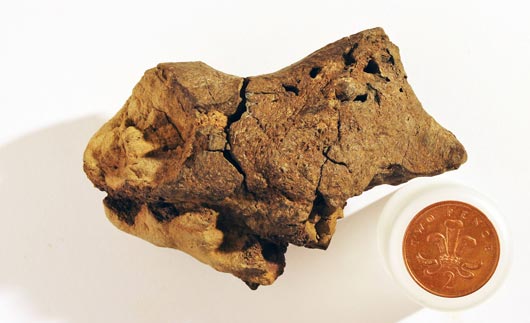
A small pebble from the foreshore at Bexhill-on-Sea is actually part of the fossilised brain of a dinosaur.
Picture credit: Jamie Hiscocks/Press Association
The Brain from an Iguanodontid
Although the genus cannot be identified, the researchers which include Dr David Norman and Dr Alex Liu (Cambridge University), are confident that this 133-million-year-old (Lower Cretaceous) fossil, came from a large ornithopod, an iguanodontid. Fossils of these herbivorous dinosaurs are known from the strata that is exposed in this area and the brain displays distinct similarities to modern-day archosaurs, the closest living relatives to the Dinosauria, crocodiles and birds.
For models and replicas of iguanodontids and other dinosaurs: CollectA Age of Dinosaurs Popular Figures and Models.
The Fossil Placed in the Correct Anatomical Position Within the Skull of an Iguanodontid
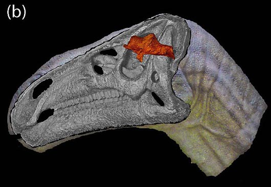
The location of the fossil within the skull of the iguanodontid.
Picture credit: Oxford University and Cambridge University
Meningeal Blood Vessels Identified
Scanning microscopy undertaken by colleagues at the University of Western Australia and three-dimensional animations provided by the University of Manchester reveal the meninges, the tough tissues surrounding the actual brain, as well as tiny capillaries and portions of the adjacent soft tissues representing the cortex. The researchers describe these details as “mineralised ghosts”.
An Image from the Three-Dimensional Animation Provided by the University of Manchester
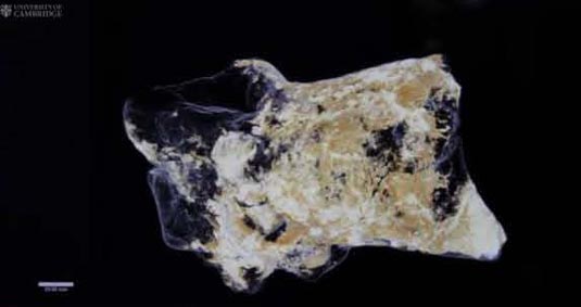
A still from the Manchester University animation which shows the fine detail on the fossil’s exterior.
Picture credit: University of Cambridge/University of Manchester
This remarkable research, a first for palaeontologists who specialise in studying the Dinosauria, has been published in a Special Publication of the Geological Society of London. The paper has been published in tribute to Professor Martin Brasier (Oxford University), who sadly died in 2014 following a road traffic accident. Professor Brasier had coordinated the research alongside Dr David Norman. One of the co-authors of the paper Dr Alex Liu was a PhD student of Professor Brasier at the time the research project was started.
Dr Liu, explained the importance of this study:
“The chances of preserving brain tissue are incredibly small, so the discovery of this specimen is astonishing.”
A Dead Dinosaur with its Head Stuck Upside Down in Stagnant Water
According to the researchers, the reason this particular piece of brain tissue has been so well-preserved is that the dinosaur’s brain was essentially ‘pickled’ in a highly acidic and low-oxygen body of water. Shortly after this dinosaur died, the corpse ended up in a shallow, stagnant pool, perhaps a bog or part of a swamp. The skull ended up buried in the fine sediment at the bottom and this allowed the soft tissue structures to be mineralised before they began to decay. This permitted the high degree of detail to be preserved.
A Magnified View of the Exterior of the Specimen
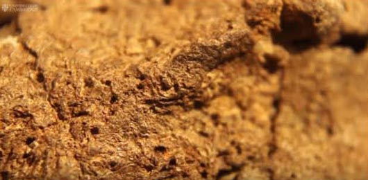
A close view of the fine structures preserved on the fossil material.
Picture credit: Oxford University and Cambridge University
Dr Norman commented:
“What we think happened is that this particular dinosaur died in or near a body of water, and its head ended up partially buried in the sediment at the bottom. Since the water had little oxygen and was very acidic, the soft tissues of the brain were likely preserved and cast before the rest of its body was buried in the sediment.”
In collaboration with researchers from the University of Western Australia, the scientists employed scanning electron microscopy in order to identify the tough membranes, or meninges, that surrounded the brain itself, as well as strands of collagen and blood vessels.
A Highly Magnified Portion of the Brain Showing the Meningeal Blood Vessels

Tubular structures identified on the exterior of the brain interpreted as representing meningeal blood vessels.
Picture credit: Oxford University and Cambridge University
Bird Brains and the Brains of Crocodilians
Such is the exceptional preservation of the specimen that the scientists have been able to identify mineralised structures that represent tissues from the cortex (the outer layer of neural tissue of the brain), along with delicate, tiny capillaries. These preserved tissues, especially the meninges are homologous to the brain tissues of extant Aves and crocodiles, close relatives of dinosaurs.
Scanning Electron Microscopy Reveals Remarkable Details
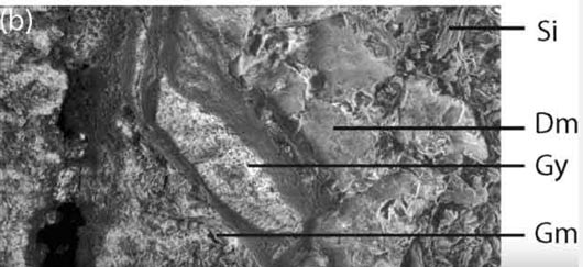
The scanning electron microscope images revealed details of the internal structure of the outermost surface of the brain.
Picture credit: Oxford University and Cambridge University
The picture above shows a highly magnified section of the fossil, sinuses as well as the tough and fibrous tissues protecting the actual brain can be made out. Could “Dm” represent the Dura mater, the tough membrane that helps to protect the brain?
A Typical Reptilian Brain
Reptile brains (and bird brains for that matter), are shaped very differently from our own brain with its substantial cerebellum. In most reptile brains, the brain is sausage-shaped, surrounded by a dense region of blood vessels and thin-walled vascular chambers (sinuses), the provide a blood drainage system. The reptile brain only occupies about fifty percent of the actual cranial cavity.
In contrast, the tissue in the fossilised brain appears to have been pressed directly against the skull, raising the possibility that some dinosaurs had large brains, with a greater volume which filled much more of the cranial cavity. With this one specimen to study, the researchers urge caution against drawing too many conclusions as to the intelligence of dinosaurs. It is likely that post-mortem, the brain got pressed against the bony roof of the cranial cavity. It is difficult to infer brain size based on the data available.
Not Sure How Big the Brain Was
Dr Norman added:
“As we can’t see the lobes of the brain itself, we can’t say for sure how big this dinosaur’s brain was. Of course, it’s entirely possible that dinosaurs had bigger brains than we give them credit for, but we can’t tell from this specimen alone. What’s truly remarkable is that conditions were just right in order to allow preservation of the brain tissue, hopefully this is the first of many such discoveries.”
A Dinosaur Brain
Fossil hunter Jamie Hiscocks, the finder of the specimen back in 2004 said:
“I have always believed I had something special. I noticed there was something odd about the preservation, and soft tissue preservation did go through my mind. Martin realised its potential significance right at the beginning, but it wasn’t until years later that its true significance came to be realised.”
Everything Dinosaur acknowledges the help of Cambridge University in the compilation of this article.
The scientific paper:
Martin D. Brasier et al.’ “Remarkable Preservation of Brain Tissues in an Early Cretaceous Iguanodontian Dinosaur.” Earth System Evolution and Early Life: A Celebration of the Work of Martin Brasier. Geological Society, London, Special Publications.
Visit Everything Dinosaur’s website: Everything Dinosaur.

Leave A Comment