Pictures of fossils, fossil hunting trips, fossil sites and photographs relating to fossil hunting and fossil finds.
3
05, 2018
The First Beak Under the Noses of Scientists
Inspiring Ichthyornis – Top of the Pecking Order
As a very young boy, I remember eagerly striving to complete my Brooke Bond “Prehistoric Animals” card collection. This was a set of fifty cards to collect, given away free with packets of tea. One of the cards featured a pair of toothed, prehistoric birds, a large, reddish coloured Hesperornis which was being mobbed by a couple of tern-like birds, this was my first introduction to Ichthyornis. Perhaps, the first time that I realised that birds (at least primitive, toothed birds), lived alongside dinosaurs.
How wonderful to read this week that Ichthyornis, thanks to a pieced together three-dimensional skull, may be providing palaeontologists with fresh insights into avian evolution. The Hesperornis/Ichthyornis picture card may have been burned into my conscience long ago, but it is refreshing to think that this ancient bird may represent a pivotal moment in the transition from dinosaurs to modern-day birds and its significance has only just come to light. A team of international scientists have published a paper proposing that Ichthyornis may have had one of the first, true bird-like beaks.
The Brooke Bond Picture Card
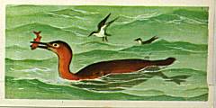
Picture credit: Everything Dinosaur/Brooke Bond
Toothy Bird with the Beginnings of a Beak
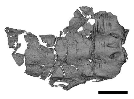
Writing in the journal “Nature”, researchers report on the analysis of beautifully preserved three-dimensional Ichthyornis (I. dispar) fossil skull that is providing new evidence on the evolution of the avian head and how the skull and beaks of birds evolved from their dinosaurian ancestors.
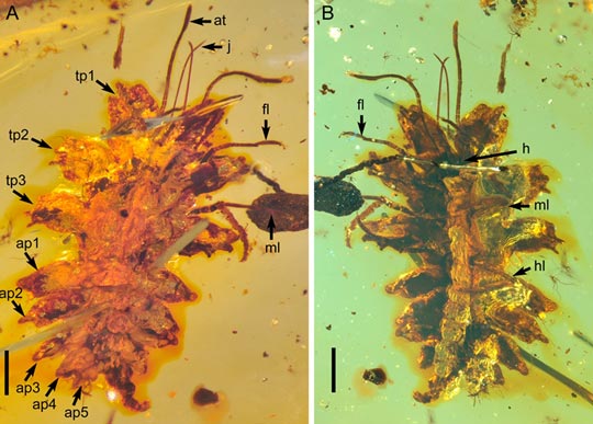
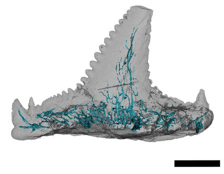
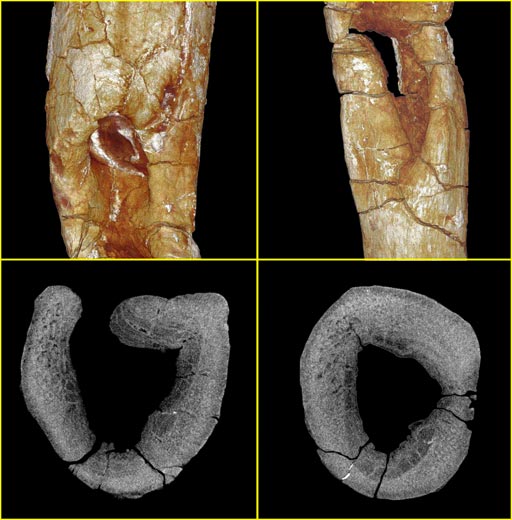
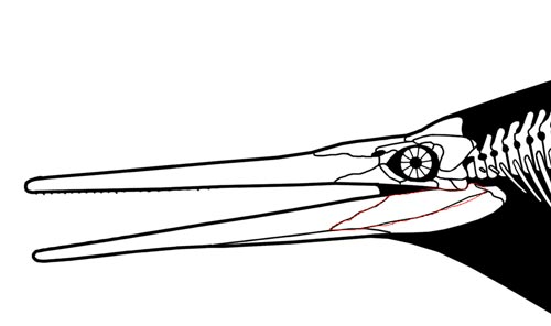
A Three-Dimensional Image of Ichthyornis Skull Material Indicates the Tip of the Premaxillary Formed the First Beak
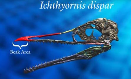
Picture credit: Yale Office of Public Affairs and Communications
Ichthyornis dispar
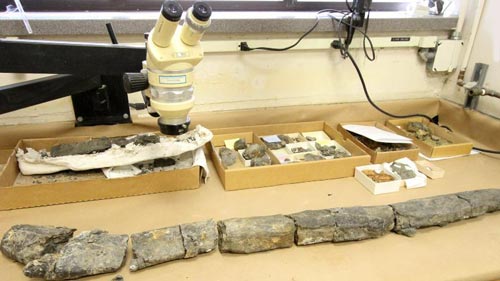
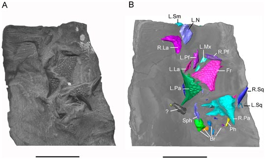
Known from fragmentary fossils from Kansas and named back in 1872 by Yale University’s Othniel Charles Marsh, it seems fitting that this new study into one of the first toothed birds described, has been led by scientists from Yale University. Working in conjunction with colleagues from the University of Kansas, Fort Hays State University, Alabama Museum of Natural History and the McWane Science Centre (Alabama), the team report on new specimens with three-dimensional cranial remains, including one example of a complete skull and two previously overlooked cranial elements that were part of the original Yale specimen examined by Marsh.
Using CT scans and sophisticated computer modelling, individual skull and jaw bones were scanned and reproduced in three-dimensions. This allowed a complete skull to be constructed revealing new details about the transition from dinosaur skull to a more modern bird skull.
The Transition from Dinosaur Skull to Bird Skull
Yale University palaeontologist and lead author of the study Bhart-Anjan Bhullar commented:
“Right under our noses this whole time was an amazing, transitional bird. It has a modern-looking brain along with a remarkably dinosaurian jaw muscle configuration.”
Ichthyornis is part of the biota of the Western Interior Seaway, a shallow sea that split North America in two during the Late Cretaceous. It has been regarded as an early version of a tern or gull, but its size is unknown as the few fossils found represent individuals of different sizes, however, it probably had a wingspan of no more than sixty centimetres, making Ichthyornis slightly smaller than today’s Common Tern (Sterna hirundo), a bird which fills the same ecological niche as the Mesozoic Ichthyornis.
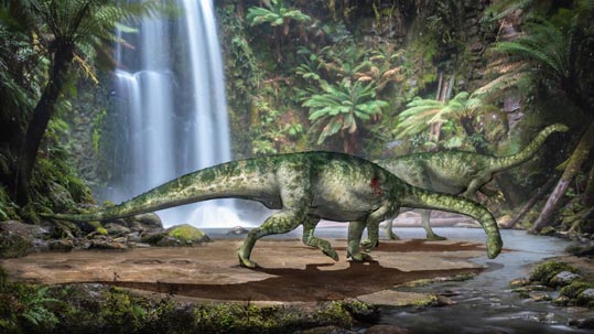
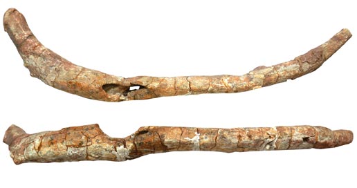
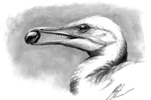
Using the Latest Research, a New Reconstruction of Ichthyornis dispar was Produced

Picture credit: Yale Office of Public Affairs and Communications
Ichthyornis – The Evolution of a Beak
Having built a three-dimensional model of the skull and jaw bones, the researchers were able to note that the premaxillary bone in the upper jaw had become elongated and this, working in conjunction with a keratinous tip on the lower jaw formed the first “proto-beak”. Ichthyornis dispar shows scientists what the first type of bird beak looked like. This beak may have evolved as the function of the hands was increasingly limited as they were adapted to form a more effective wing.
The grasping hands of the maniraptoran dinosaurs were no longer able to grasp and manipulate objects so the jaws had to take on an additional function, secondary to their main function – dispatching and consuming prey.
The Beak of Ichthyornis
Picture credit: Yale Office of Public Affairs and Communications
Although maniraptoran dinosaurs may not have been able to pronate their hands like us and they lacked an opposable thumb, as forelimbs and hands evolved into wings, so the jaws took over the function of the digits and manus.
Bhart-Anjan Bhullar stated:
“The first beak was a horn-covered pincer tip at the end of the jaw. The remainder of the jaw was filled with teeth. At its origin, the beak was a precision grasping mechanism that served as a surrogate hand as the hands transformed into wings.”
The research team conducted its analysis using CT-scan technology, combined with specimens from the Yale Peabody Museum of Natural History; the Sternberg Museum of Natural History in Hays, Kansas, the Alabama Museum of Natural History; the University of Kansas Biodiversity Institute and the Black Hills Institute of Geological Research (South Dakota).
Bird Beaks versus Bird-hipped Dinosaur Beaks
The modern bird beak is a unique organ amongst vertebrates, although notably most derived ornithischian (bird-hipped) dinosaurs possessed a beak, formed from the unique predentary bone in the lower jaw and a roughened, extension of the premaxilla (or the rostral in the case of ceratopsians), in the upper jaw, which allowed the attachment of a keratinous tip which in conjunction formed the beak-like structure – believed to be an adaptation to assist with cropping vegetation.
This study of Ichthyornis suggests that the first bird beak was not the long organ seen in modern birds, but a little pincer tip to grasp and manipulate objects.
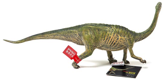
A Chasmosaurine Ceratopsian with the Roughened Rostral and the Predentary Forming a Plant-cropping Beak
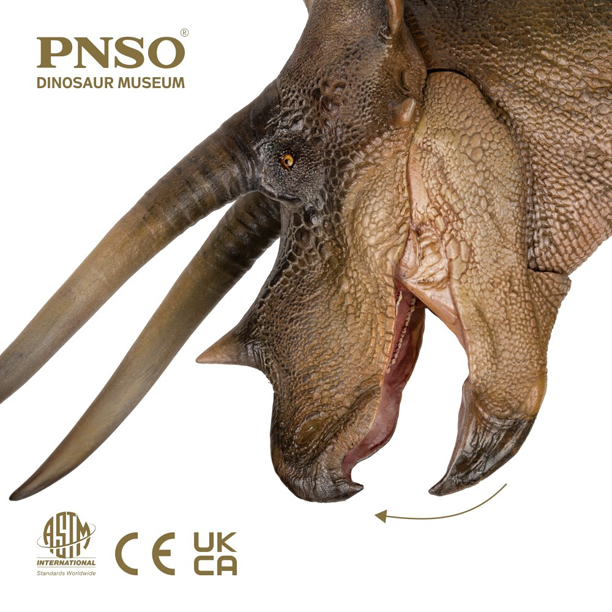
The picture (above) shows a ceratopsian figure from the PNSO model range.
To view these figures: PNSO Age of Dinosaurs Models.
Fresh Insight into the Evolution of Extant Bird Skulls
The scientists conclude that their study offers new insights into how modern birds’ skulls formed. Along with its transitional beak, Ichthyornis dispar had a brain similar to that seen in extant birds but a temporal region of the skull that was reminiscent of a dinosaur. This suggests that during the evolution of Aves, the brain transformed first, possibly to adapt to a volant (aerial) lifestyle, whilst the remainder of the skull retained the ancestral features associated with the Dinosauria.
Ichthyornis retained a large adductor chamber bounded at the top by substantial bony remnants of the ancestral reptilian upper temporal fenestra (hole in the skull). This combination of features indicates that important attributes of the avian brain and palate evolved before the reduction of jaw musculature and the full transformation of the beak.
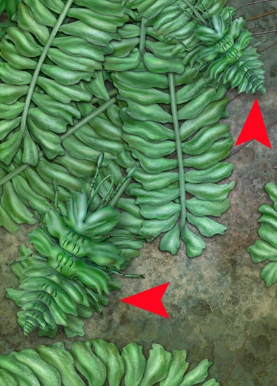
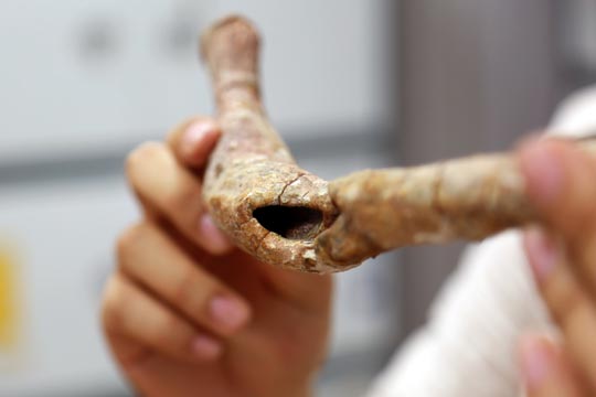
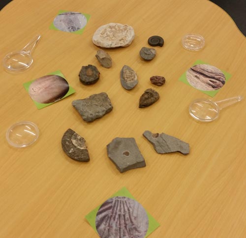
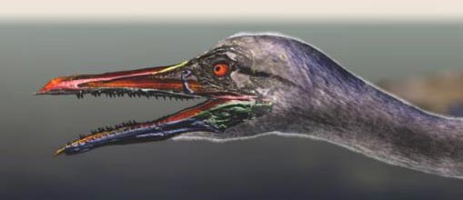
The Beak of Ichthyornis Grasping a Mollusc
Picture credit: Michael Hanson/Bhart-Anjan Bhullar
I may never have completed my Brooke Bond card collection, but at least, thanks to this new Ichthyornis study, our understanding of the evolution of the beak in birds is more complete.
Visit the Everything Dinosaur website: Everything Dinosaur.