Pictures of fossils, fossil hunting trips, fossil sites and photographs relating to fossil hunting and fossil finds.
7
02, 2019
New Defensive Dicraeosaurids – Forward Facing Spikes Deter Predators
Bajadasaurus pronuspinax – Sauropod Defences
A newly described sauropod from northern Patagonia (Argentina), has provided palaeontologists with evidence to help explain why some of these long-necked dinosaurs evolved long, paired spines on the necks. These features may have had a primary role as defensive structures helping to deter attacks from theropod predators. The dinosaur has been named Bajadasaurus pronuspinax and it has been assigned to the Dicraeosauridae family, a sister family to the Diplodocidae within the Sauropoda. Dicraeosaurids are characterised by having relatively shorter necks and distinctive vertebrae which had long, paired neural spines. The function of these spines has long been debated. They have been interpreted as playing a role in visual communication, sexual display and thermoregulation, however, this newly described dinosaur suggests that within this family of long-necked dinosaurs they evolved as a form of defence.
Illustrating Bajadasaurus pronuspinax and the Fossil Find Location
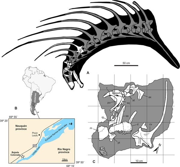
Picture credit: Gallina et al published in Scientific Reports
The image (above), shows a skeletal reconstruction of the head and neck of Bajadasaurus (A), with the preserved fossil material shown in white. On the right of the image is a location map (B), showing the site of the fossil find, close to the Ezequiel Ramos Mexía lake in Neuquén Province, Argentina. A line drawing is provided (C), that shows the association and the location of the fossils found at the dig site.
Interpreting Fossils One Cervical Vertebra at a Time
The authors of the scientific paper, propose that the elongated neural spines of this dinosaur always faced forward, presenting a formidable obstacle for any meat-eating dinosaur wanting to attack the animal’s neck. However, it is worth noting that if the image (above), is studied, the theory of Bajadasaurus having a neck topped with defensive spikes, like some sort of Victorian railings is based on the discovery of a single neck bone, in the skeletal illustration placed in the position of the fifth cervical vertebra. The appearance of B. pronuspinax is inferred by comparing these fossils to the better-known Amargasaurus (A. cazaui). Until more fossils are found the appearance of Bajadasaurus and the orientation of those neural spines can only be speculated.
A Model of the Dicraeosaurid Amargasaurus

Lead-author of the study, Pablo Gallina and his colleagues, propose that these neural spines may have been covered with keratin and therefore much longer than the spines themselves. The extent of the neural spines, the length of the keratin sheaths that covered them and the direction they pointed in, remains unknown. Until more fossils of Bajadasaurus are found, those elongated neural spines remain a mystery.
Naming a New Dinosaur
That one cervical spine forms the basis for the species epithet. The genus honours Bajada (Spanish for downhill), a reference to the fossil find location – Bajada Colorada. The species name means “bent over, forward spines”, we shall see if more fossil discoveries reaffirm this interpretation. Importantly, the fossil material assigned to Bajadasaurus includes much of the skull, thanks to these fossils, the palatal bones, the braincase and a nearly complete left dentary, palaeontologists have a much better idea about the size and morphology of dicraeosaurid dinosaur skulls.
Skull Material Associated with Bajadasaurus pronuspinax and Line Drawing
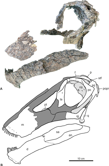
Picture credit: Gallina et al published in Scientific Reports
The skull is quite small for a sauropod, dicraeosaurids described to date were not as big as some of their diplodocid cousins. Size estimates range from around 10 to 13 metres in length. The size of Bajadasaurus is unknown, but based on these fossils, it is likely that this dinosaur was within this size range too. The orbits are quite large and their position on the top of the skull suggests that when this dinosaur had its head down and it was feeding, it was capable of seeing ahead (forward-directed, stereoscopic vision).
Comparing Bajadasaurus to the Geologically Younger Amargasaurus
The strata of the Bajada Colorada Formation represent sediments laid down at the very beginning of the Cretaceous (Lower Cretaceous, Berriasian/Valanginian faunal stages). Bajadasaurus roamed Patagonia some 140 million years ago. Amargasaurus, lived in the same part of South American but around fifteen million years later.
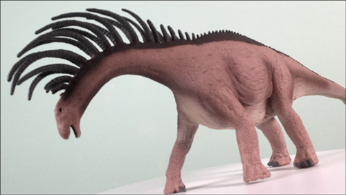
A Model of the Sauropod Dinosaur Bajadasaurus
Picture credit: Everything Dinosaur
The picture (above) shows a CollectA Deluxe Bajadasaurus dinosaur model.
To view this model range: CollectA Deluxe Age of Dinosaurs Models.
The researchers suggest that the temporal difference between Bajadasaurus and Amargasaurus, supports the idea that the development of an array of defensive spines was likely adaptive over a long time period. How effective these spines may have been against predators, is once again, open to speculation. However, the presence of elongated neural spines would have given the impression of a larger animal with a thicker neck. To a hungry, carnivorous dinosaur the appearance of a bigger more robust adversary may have been enough of a deterrent.
The scientific paper: “A New Long-spined Dinosaur from Patagonia Sheds Light on Sauropod Defence System” by Pablo A. Gallina, Sebastián Apesteguía, Juan I. Canale and Alejandro Haluza published as an open access article in the journal “Scientific Reports”.
Visit the Everything Dinosaur website: Everything Dinosaur.