Fossil finds, new dinosaur discoveries, news and views from the world of palaeontology and other Earth sciences.
21
12, 2022
An Amazing Fossil – Dinosaur Eating a Mammal
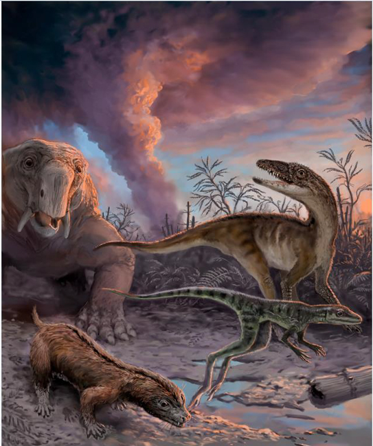
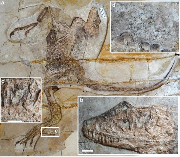
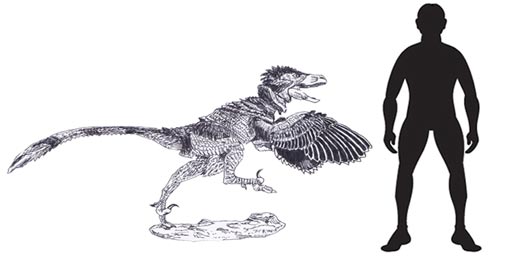
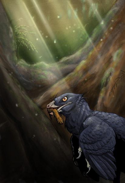
The first, definitive proof of a dinosaur eating a mammal has been found. A foot of a tiny, mouse-sized mammal has been discovered inside the body cavity of the feathered theropod Microraptor (M. zhaoianus). Previously, other Microraptor specimens from Lower Cretaceous rocks of northern China had revealed the fossilised remains of a fish, a primitive bird and a lizard associated with the body cavity. Palaeontologists now know that this crow-sized predator also ate mammals. This is the first record of a dinosaur consuming a mammal.
Mammal Foot Found Inside Ribcage
A new study led by Dr David Hone (Queen Mary University of London), published in the academic “Journal of Vertebrate Paleontology”, documents the first known incident of a dinosaur having eaten a mammal.
Microraptor is a genus of small, dromaeosaurid which lived in the forests of northern China around 120 million years ago (Early Cretaceous). The remarkable fossils found in Liaoning Province have enabled palaeontologists to build up a detailed picture of life in these ancient, dinosaur-dominated forests.
Researchers have also identified a wide variety of mammals and mammaliamorphs that co-existed with the dinosaurs and pterosaurs. Together these creatures make up a diverse ecosystem known as the Jehol biota
To read Everything Dinosaur’s blog post from 2021 describing the remarkable diversity of vertebrates associated with the Jehol biota: The Jehol Biota.
Microraptor had long feathers on its arms and legs and was, very probably arboreal, gliding from tree to tree, hunting out small animals to eat.
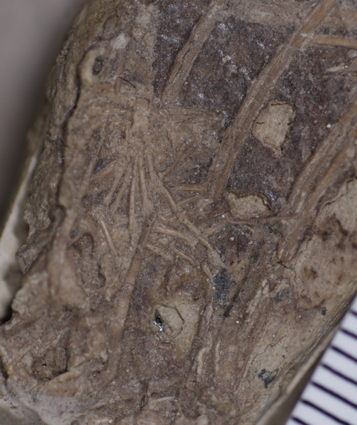
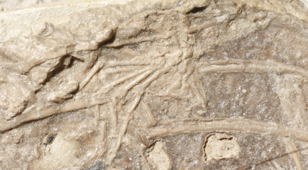
Spotting the Fossilised Foot
The Microraptor specimen was first described twenty-two years ago, but the preserved remains of the tiny foot had been overlooked. Professor Hans Larsson of McGill University in Montreal spotted what others had missed – the remains of another animal inside the Microraptor’s rib cage. In collaboration with Dr Hone, and colleagues from Canada, China and the USA, a paper describing this remarkable discovery has now been published.

Dinosaur Eating a Mammal
The mammal foot is almost complete and belonged to a very small animal, approximately the size of a modern house mouse. Examination of the bones suggest that it was one that predominantly lived on the ground and was not well adapted for climbing trees, making it an interesting prey choice for the mainly arboreal Microraptor.
Previous studies have revealed other Microraptor specimens containing the remains of a bird, a lizard and a fish. This specimen of the species Microraptor zhaoianus demonstrates that Microraptor also consumed small mammals. This little feathered dinosaur was a generalist, consuming a wide variety of prey.
It is not certain if the dromaeosaurids in question had directly preyed upon and eaten these animals or found them already dead and had scavenged them (or a mixture of both) but the mammal at least falls into the range of typical prey size predicated for a predator the size of Microraptor.
Dr Hone’s co-authors on the paper include Dr Alex Dececchi, Mount Marty College (USA), Dr Corwin Sullivan at the Department of Biological Sciences, University of Alberta, and Professor Xu Xing at the Institute of Vertebrate Palaeontology and Palaeoanthropology, Beijing.
A Significant Fossil Discovery
Commenting on the significance of this fossil discovery, Dr David Hone stated:
“It’s so rare to find examples of food inside dinosaurs so every example is really important as it gives direct evidence of what they were eating.“
Dr Hone from the University’s School of Biological and Behavioural Sciences added:
“While this mammal would absolutely not have been a human ancestor, we can look back at some of our ancient relatives being a meal for hungry dinosaurs. This study paints a picture of a fascinating moment in time – the first record of a dinosaur eating a mammal – even if it isn’t quite as frightening as anything in Jurassic Park.”
Co-author of the study, Dr Alex Dececchi, from Mount Marty College, commented:
“The great thing is that, like your housecat which was about the same size, Microraptor would have been an easy animal to live with but a terror if it got out as it would hunt everything from the birds at your feeder to the mice in your hedge or the fish in your pond.”
Everything Dinosaur acknowledges the assistance of a media release supplied by Dr David Hone in the compilation of this article.
The scientific paper: “Generalist diet of Microraptor zhaoianus included mammals” by Hone, D.W.E., Dececchi, T.A., Sullivan, C., Xu, X. and Larsson, H.C.E. published in the Journal of Vertebrate Paleontology.
Correction
This is not the first recorded incidence of a dinosaur consuming a mammal. The press release, although provided by the appropriate authorities, had failed to recognise evidence cited in an earlier scientific paper.