All about dinosaurs, fossils and prehistoric animals by Everything Dinosaur team members.

Fossil finds, new dinosaur discoveries, news and views from the world of palaeontology and other Earth sciences.

22 11, 2016

Bird Melanosome Stories Flocking Together as New Research is Published

By |2024-05-06T15:36:14+01:00November 22nd, 2016|Categories: Dinosaur and Prehistoric Animal News Stories, Dinosaur Fans, Main Page|0 Comments

Keratin and Melanosomes Preserved in Eoconfuciusornis

A team of international scientists led by researchers from the Chinese Academy of Sciences in collaboration with colleagues from North Carolina State University and Linyi University (Shandong, China), have reported finding the oldest fossil evidence of beta-keratin and melanosome preservation in the feathers of a 130-million-year-old primitive bird from the famous Early Cretaceous Jehol Biota of north-eastern China.

Keratin and Melanosomes Preserved in Eoconfuciusornis

Writing in the journal “The Proceedings of the National Academy of Sciences (United States)”, this new study extends the time frame in which evidence of original organic molecules may be preserved and has provided new data on how to distinguish melanosome structures from similarly shaped microbes.  This is the second article related to the melanosomes having been identified in ancient birds that Everything Dinosaur has published the week.  The earlier article related to a bird fossil identified as a member of the Enantiornithes clade excavated from the Lower Cretaceous strata (Jiufotang Formation), that showed evidence of iridescence in its feathers.*  It’s almost as if stories relating to melanosome structures in birds are flocking together!

The Lower Cretaceous rocks exposed in the Chinese provinces of Hebei and Liaoning have provided palaeontologists with a huge number of vertebrate fossils to study.  The fine-grained strata, laid down in a lacustrine (lake environment), surrounding by active volcanoes provided ideal body fossil preservation conditions and many specimens have revealed exquisite details.  One of the most important group of fossils associated with these deposits are the many different types of bird fossil that have been found.   This new study was carried out on the fossilised remains of one such primitive bird, a specimen of Eoconfuciusornis.

Scanning Transmission Microscopy

Scanning transmission microscopy was undertaken to identify structures within the fossil and the surrounding matrix, chemical analysis including the use of antibodies that react to specific proteins, helped pinpoint the identity of the structures that had been found.

The Fossilised Remains of Eoconfuciusornis Used in the Research
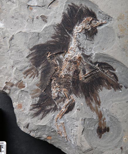
Eoconfuciusornis fossil bird.

The fossilised remains of a Lower Cretaceous bird from China (Eoconfuciusornis).

Picture credit: Dr Xiaoli Wang (Linyi University)

For models and replicas of Chinese prehistoric animals including confuciusornithids: Dinosaur Models and Figures (PNSO).

What Sort of Bird was Eoconfuciusornis?

Eoconfuciusornis was a crow-sized member of the primitive bird group the confuciusornithids that thrived in China during the Early Cretaceous.  It was one of the first birds to possess a pygostyle (the last five tail vertebrae fused together) and to have a keratinous beak lacking teeth, features found in all modern birds (Neornithes).

Previous studies had striven to prove that tiny, round or sausage-shaped structures preserved in association with fossilised remains were melanosomes, organelles found in animal cells that contains the light-absorbing pigment melanin responsible for colour and protection from harmful rays emitted from the sun.

However, although melanosome-like structures had been discovered in a number of specimens, there was an element of doubt as to the authenticity of the research findings.  It was difficult to definitively proved that these structures were not just microbes that had become associated with the feathers during decomposition and fossilisation.

Studying Melanosomes

Mary Schweitzer, a leader in the field of identifying traces of organic materials from within the fossil record and a co-author of this research paper explained the problem:

“If these small bodies are melanosomes, they should be embedded in a keratinous matrix, since feathers contain beta-keratin.  If we couldn’t find the keratin, then those structures could as easily be microbes, or a mix of microbes and melanosomes, in either case, predictions of dinosaur shading would not be accurate.”

The scientists used both scanning and transmission electron microscopy to map minute details of the feather’s surface and its internal structure.  They also utilised immunogold tagging, a technique which involves gold, non-reactive particles being attached to antibodies that bind to particular proteins in order to make them visible in electron microscopy.  This helped to show that the fine filaments within the feathers were keratin.

An Illustration of a Typical Confuciusornithid Bird of the Lower Cretaceous (Confuciusornis sanctus)
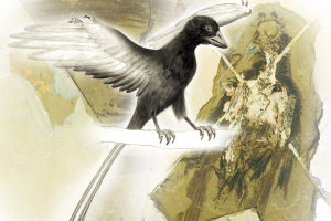
Confuciusornis sanctus.

Confuciusornis sanctus a primitive bird but it had a beak.

Picture credit: University of Manchester

Mapping Chemical Elements

Further tests were carried out, including one which involved the mapping of the presence of copper and sulphur in association with the feathers when viewed under high resolution.  Sulphur was broadly distributed, reflecting its presence in both melanin molecules and keratin.  However, the distribution of copper (only found in modern melanosomes and not part of keratin), was only observed in the fossil melanosome structures.  The data produced supports the identity of melanosomes and indicates that these structures are not the result of contamination or microbial action.

Fellow author, Yanhong Pan (Chinese Academy of Sciences) stated:

“This study is the first to demonstrate evidence for both keratin and melanosomes, using structural, chemical and molecular methods.  These methods have the potential to help us understand, on the molecular level, how and why feathers evolved in these lineages.”

If melanosomes can be positively identified within the fossil record and as the shape of melanosomes can provide an indication of animal colouration, then palaeontologists may be able to hypothesise with greater confidence the colouration of extinct animals including dinosaurs and primitive birds.

* To read Everything Dinosaur’s recently published article on the study of a member of the Enantiornithes clade that shows evidence of iridescence in preserved feathers: A Prehistoric Bird with Bling.

The scientific paper: “Molecular Evidence of Keratin and Melanosomes in Feathers of the Early Cretaceous Bird Eoconfuciusornis”, published in the Proceedings of the National Academy of Sciences (United States of America).

The Everything Dinosaur website: Everything Dinosaur.
21 11, 2016

Ginkgo “Living Fossil” Has Remarkable Genome Sequenced

By |2023-05-09T12:58:12+01:00November 21st, 2016|Categories: Dinosaur and Prehistoric Animal News Stories, Dinosaur Fans, Main Page, Photos/Pictures of Fossils|0 Comments

The Genome of the Ginkgo Reveals Secret of Success

A team of international scientists writing in the open-access journal “GigaScience”, have published a paper that maps the genome of a “living fossil” – the Ginkgo tree.

The extant Ginkgo (Ginkgo biloba), is the only representative of a division of the Plantae Kingdom called the Ginkgophyta.  Native to China, this slow-growing tree with its distinctive fan-shaped leaves has a fossil record that dates back to the Permian.  Also called the Maidenhair tree, Ginkgo biloba is the only living species of a once diverse and widespread group that thrived during the Mesozoic.  The evolutionary origins of the Ginkgophyta are unclear, with the huge genome of G. biloba mapped, it may be possible to trace the ancestry of this enigmatic plant.

The Genome of the “Living Fossil” Ginkgo Biloba has been Mapped

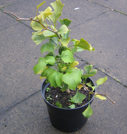
A Ginkgo biloba tree.

A small Ginkgo tree (G. biloba). The genome of Ginkgo biloba has been mapped.

Picture credit: Everything Dinosaur

Visit Everything Dinosaur’s website: Everything Dinosaur.

What is a Genome?

A genome is a complete set of genetic instructions required to build an organism, allow it to grow, develop and function properly.  The instructions are comprised of DNA (Deoxyribonucleic acid), which itself consists of four nucleotide bases adenine, thymine, cytosine and guanine (plus other materials that make up the DNA molecule such as phosphate and sugars).

The DNA is contained in chromosomes, which are found in the nucleus of most cells. The four nucleotides are arranged in pairs, adenine binds to thymine and cytosine unites with guanine.  The gene is the unit of inheritance, sections of the DNA consisting of an arrangement of the four base pairs make up a single gene and it is this gene that provides the instructions to build and maintain the organism.

Each organism has a unique genome and the length of these genetic instructions varies.  For example, the human genome is comprised of around 3.2 billion base pairs, if the genome of H. sapiens was printed out and put into paperback books, the stack of books would be over sixty metres tall, about as high as the monument to the Great Fire of London adjacent to London Bridge.

The Ginkgo Tree Has a Much Bigger Genome

However, the Ginkgo has a much bigger genome, consisting of approximately 10.6 billion base pairs.  If the genome of G. biloba was printed into paperback form, the stack of books would reach over 200 metres high, that’s as tall as the giant cooling towers of the Fidlers Ferry Power station that dominates the Cheshire landscape.

The Human Genome Compared to the Much Larger Genome of Ginkgo biloba

Human genome compared to the Ginkgo genome.

If the human genome and the Ginkgo genome were printed as paperbacks, the tower of books representing the Ginkgo genome would be more than three times higher.

Picture credit: Everything Dinosaur

In the picture above, the human genome if printed out into paperback books, would make a stack some 62 metres tall.  The much larger genome of G. biloba would make a tower of books over 200 metres high.  A Brachiosaurus is shown for scale.

The Extraordinary Biology of G. biloba

The research team, which included members of the Chinese Academy of Sciences and scientists from Zhejiang University, are trying to find out why individual trees can live so long and how they overcome insect and fungi attacks.  The Ginkgo genome mapping study is part of a much larger research project studying plant defences against pathogens.  This investigation will help palaeobotanists to better understand the evolutionary history of the Ginkgophyta and how they relate (phylogeny) to other plant groups.

Professor Yunpeng Zhao (Zhejiang University), one of the authors of the scientific paper commented:

“Ginkgo represents one of the five living groups of seed plants, and has no living relatives.  Such a genome fills a major phylogenetic gap of land plants, and provides key genetic resources to address evolutionary questions like phylogenetic relationships of gymnosperm lineages, evolution of genome and genes in land plants, innovation of developmental traits, evolution of sex as well as history of demography and distribution, resistance and conservation of the Ginkgo.”

Fossilised Ginkgo Leaves (Jurassic)

The carbonised fossil leaves of a Jurassic Ginkgo tree.

Ginkgo fossil leaves.

Picture credit: Everything Dinosaur

A Very Resilient Plant

The resilience of the Ginkgo has fascinated botanists.  It has survived dramatic climate change including mini Ice Ages in its native China and Ginkgo trees in the Japanese city of Hiroshima were one of the few organisms to survive the atom bomb blast and resulting radiation, the oldest recorded individual tree germinated back in the Bronze Age.

Ginkgo trees are also able to fend off attacks from insects, bacteria and fungi.  The leaves contain an arsenal of complex chemicals that provide an effective defence.  To better understand the Ginkgo’s defensive systems, the authors analysed the repertoire of genes present in the genome that are known to play a role in the defence of the plant.  An initial analysis of the tree’s more than 40,000 predicted genes showed extensive expansion of gene families that provide for a wide range of defensive mechanisms.

Genes that enable resistance against pathogens are often duplicated.  In addition, Ginkgo trees have an extra weapon in their battle against arthropods.  As well as having their own array of insecticides within their leaves, they also defend themselves against attack by releasing volatile organic compounds that specifically attract enemies of plant-eating insects.

A Living Fossil – Ginkgo biloba

The term “living fossil” is not one readily used by scientists.  It implies that a species has survived unchanged of millennia.  This is not true, the fossilised leaves preserved in Mesozoic and Cenozoic strata represent a wide variety of genera, but the tree’s basic body plan, its design has remained relatively unchanged for millions of years.

This new data, indicating that the Ginkgo has a formidable array of weaponry to keep it safe from harm, defence measures that seem to have come about as the genes were duplicated, might be linked to the plant’s remarkable resilience.  By undertaking a study of the Ginkgo and its genome, scientists may find useful pointers to assist them in helping to produce more robust crops that can resist diseases and pests.

The CollectA Ginkgo biloba Model

CollectA added a twenty-five centimetre high model of a Ginkgo tree into their “Prehistoric Life Collection” a couple of years ago.  This replica works well with the 1:20 scale range of prehistoric animal models and is very useful when creating realistic prehistoric scenes and dioramas.

The CollectA Prehistoric Life Ginkgo biloba Model

 CollectA Ginkgo biloba model.

The CollectA Ginkgo biloba tree model.

To view the CollectA Ginkgo biloba replica and the other scale models in this range: CollectA Deluxe Prehistoric Animals and Models of Prehistoric Trees.

20 11, 2016

Just When Did the Dinosaurs Dominate the Land?

By |2023-05-09T12:45:55+01:00November 20th, 2016|Categories: Dinosaur and Prehistoric Animal News Stories, Dinosaur Fans, Main Page, Palaeontological articles|0 Comments

Ixalerpeton polesinensis and Buriolestes schultzi Co-existed

New fossil evidence suggests that the rise of the Dinosauria was more gradual than previously thought.  Many people’s perception of the dinosaurs is that they are all super-sized monsters, dominating life on land and rapidly out competing the other, more primitive Triassic reptiles.  Scientists writing in the journal “Current Biology” challenge this view, as they describe the discovery in the same rock formation of an early dinosaur and a lagerpetid, a member of a group of animals that are recognised as precursors of dinosaurs.

This is the first time that a dinosaur and a dinosaur precursor have been found together, indicating that true members of the Dinosauria Order and their near relatives co-existed.

The Skull of the Dinosaur (Buriolestes schultzi) in Situ

Buriolestes skull at the dig site.

The skull of the sauropodomorph Buriolestes.

Picture credit: Cabreira et al

A Sauropodomorph and a Lagerpetid

 A team of researchers including Sergio Furtado Cabreira (Museu de Ciências Naturais, Universidade Luterana do Brasil, Brazil) and Alexander Wilhelm Armin Kellner (Departamento de Geologia e Paleontologia, Museu Nacional-UFRJ, Rio de Janeiro, Brazil) have described two new species of Dinosauromorpha from Upper Triassic rocks in central, eastern Brazil (the Paraná Basin).

The fossil material represents a lagerpetid, which has been named Ixalerpeton polesinensis and a new basal sauropodomorph (Buriolestes schultzi), the remains of these animals were found in the Alemoa Member of the Santa Maria Formation, specifically, within the Hyperodapedon (rhynchosaur) Assemblage Zone which dates from the Carnian faunal stage of the Upper Triassic.

Ixalerpeton has been assigned to the family Lagerpetidae which together with the Dinosauria and other closely related archosaurs, make up the clade Dinosauromorpha.  B. schultzi has been assigned to the Sauropodomorpha, a clade of lizard-hipped dinosaurs that would evolve into the long-necked giants such as Diplodocus, Apatosaurus and Brachiosaurus.

Line Drawings and Photographs of the Fossilised Remains of the Two New Species of Dinosauromorph

Fossils and drawings of the two Dinosauromorpha.

Line drawing (A) of I. polesinensis and fossilised remains of (B-H) and line drawing (I) of B. schultzi with fossil remains (J-P).

Picture credit: Current Biology

The Lagerpetid Ixalerpeton polesinensis

The lagerpetid (Ixalerpeton) has been described from skull bones including the braincase, vertebrae, one shoulder blade, the left humerus, parts of the pelvis, a thigh bone (femur) and some lower leg bones.  This biped is estimated to have measured around half a metre in length.

The Sauropodomorph Buriolestes schultzi

The larger Buriolestes is estimated to have measured around 1.8 metres long and it has been described from a partial skull, which includes the premaxilla, maxilla and the dentary, a large number of vertebrae including posterior dorsal vertebrae and caudal bones, the left arm, parts of the pelvis and a nearly complete left hind leg.

For scale models of Triassic prehistoric animals and early dinosaurs: CollectA Deluxe Prehistoric Animal Replicas.

Etymology

Ixalerpeton polesinensis – The genus name means “leaping reptile”, whilst the trivial name references São João do Polêsine, the town where the fossils were found.

Buriolestes schultzi – The genus name means “Buriol robber”, a reference to the family name of the land owners and recognises that this dinosaur was most likely carnivorous.  The species/trivial name honours Cesar Schultz (professor of vertebrate palaeontology at the Universidade Federal do Rio Grande do Sul).

A Timeline Showing the Relationship between B. schultzi and I. polesinensis within an Overview of Early Dinosauromorphs

A timeling showing the evolutionary relationships of early dinosauromorphs.

Phylogeny of early dinosauromorphs.

Picture credit: Current Biology

Ixalerpeton polesinensis and Buriolestes schultzi

The research team are confident that further analysis of the Ixalerpeton fossil material will add to our understanding as to how the dinosaurs evolved their anatomical characteristics.  The teeth of Buriolestes indicate that this sauropodomorph, a distant ancestor of the huge herbivorous sauropods, was actually carnivorous.  This evidence supports the theory that the giant plant-eating dinosaurs such as Diplodocus were descended from small, bipedal, meat-eaters.

A Close View of the Teeth of Buriolestes Indicating that this Dinosaur was a Carnivore

The teeth of Buirolestes

A close view of the teeth of Buriolestes. The highly recurved and serrated teeth indicate that this sauropodomorph was a carnivore.

Picture credit: Cabreira et al

The Dietary Preferences of Dinosauromorphs

An analysis of the fossil teeth of these newly described archosaurs has helped scientists to assess the dietary preferences of a range of dinosauromorphs.  This research suggests that the very first dinosaurs were all meat-eaters and over time there was a move towards herbivory and an omnivorous diet within certain groups.

The Dietary Preferences of Dinosauromorphs

The diets of dinosauromorphs.

The dietary preferences of members of the Dinosauromorpha.

Picture credit: Current Biology

The diagram above shows a cladogram of dinosauromorphs and photographs of their teeth.  The top photograph shows the teeth of the dilophosaurid theropod Dracovenator (Neotheropoda), a carnivore.  Next comes a picture of the teeth of Buriolestes, assigned to the Sauropodomorpha but regarded as carnivorous by the researchers.  The third photograph features the teeth of Pampadromaeus, which coincidentally, was named by Sergio F. Cabreira, Cesar L. Schultz et al.  This little dinosaur has been assigned to the Sauropodomorpha too, it is also believed to be meat-eater.  The final set of teeth belong to the later sauropodomorph Plateosaurus, that lived some fifteen million years after Buriolestes and Pampadromaeus.  The teeth of Plateosaurus seem to be adapted to a plant-eating diet.

The hypothesis about feeding preferences advocated by the researchers in this paper is shown on the left of the cladogram, with alternative arrangements shown on the right.

Key

green coloured line = herbivory and/or omnivory

black coloured line = unknown or ambiguous

orange = carnivore (faunivory – feeding on other animals)

An Illustration of a Buriolestes Catching a Hyperodapedon whilst a Group of Ixalerpeton Scatter

A Buriolestes and Ixalerpeton illustrated.

A Buriolestes catches a rhynchosaur whilst a group of Ixalerpeton hunt for lizards and grubs in the undergrowth.

Picture credit: Oliveira Maurílio

Dinosaurs Evolved in the Southern Hemisphere

This research lends weight to the hypothesis that dinosaurs evolved in the Southern Hemisphere and that the Dinosauria were carnivorous in their basal forms, it also supports the idea that lagerpetids and early dinosaurs were contemporaries since the first stages of dinosaur evolution.

The scientific paper: “A Unique Late Triassic Dinosauromorph Assemblage Reveals Dinosaur Ancestral Anatomy and Diet” published in Current Biology.

Everything Dinosaur’s website: Everything Dinosaur.

16 11, 2016

Prehistoric Bird with “Bling” Identified in New Study

By |2024-05-06T15:31:02+01:00November 16th, 2016|Categories: Dinosaur and Prehistoric Animal News Stories, Dinosaur Fans, Main Page|0 Comments

Study Suggests Early Cretaceous Bird Had Flashy Feathers

Fossils from the Jiufotang Formation of Liaoning Province (China), have provided scientists with an extensive data set that records life in the Early Cretaceous.  The highly fossiliferous strata (referred to as a Lagerstätte), has given palaeontologists a deep understanding of the fauna of a forested habitat with many large lakes.

Early Cretaceous Bird with Bling

Numerous fossils of turtles, pterosaurs, fish, mammals and dinosaurs have been described, but perhaps the Jehol Biota’s greatest contribution to our understanding of vertebrate evolution has come from the large numbers of bird fossils that have been found.  A team of international researchers have studied the ancient feathers of one such bird and deduced that it had sparkly, iridescent feathers probably for display in a bid to attract a mate.

Ancient Prehistoric Bird Provides Evidence of Iridescent Feathers

Ventral view and line drawing of the bohaiornithid bird specimen.

Ventral view of the new bohaiornithid specimen (CUGB P1202, primary slab). A, photograph of the primary slab. B, interpretive drawing.

Picture credit: Jennifer A. Peteya (University of Akron)

A Bohaiornithid Bird with Bling

Scientists from Akron University (Ohio), the University of Texas at Austin in association with Chinese colleagues have studied the slab and counter slab of a beautifully preserved primitive bird.  Using high magnification and a non-destructive chemical technique to identify molecule signatures (Ramon spectroscopy), the team have identified melanosome structures that are associated with iridescent colouration in modern birds.

The image above shows a view of the primary slab showing the bird fossil (A), whereas (B) shows an interpretative line drawing of this enantiornithine.  The bird has been classified as a basal member of the Enantiornithes clade and the teeth in the tiny beak have led to the fossil being assigned to the Bohaiornithidae family (predatory birds).  As far as we at Everything Dinosaur are aware, no genus name has been assigned as yet.

The Rich and Diverse Early Cretaceous Forests of Northern China

The rich and diverse Daohugou Biota.

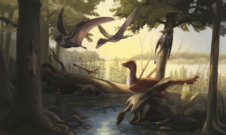
A rich and diverse environment dominated by small mammals, pterosaurs, primitive birds and feathered theropods.

Picture credit: Julia Molnar

What are Melanosomes?

Using electron microscopes to look inside the feathers of the ancient bird, the research team were able to identify strange shapes and structures.  These are melanosomes, which when the animal was alive would have contained the pigment melanin.  This light-absorbing pigment is what gives colour to human hair and animal fur, as well as to the feathers of dinosaurs and birds.  The shape of the melanosome suggests the colour.  The crown, neck, and body contour feathers retain elongate melanosome structures associated with weakly iridescent colouration in living birds, so the team have concluded that this prehistoric bird was also iridescent.

Identifying Melanosomes in a Fossil Bird

Evidence of preserved melanosomes in Early Cretaceous bird.

The right wing and body contour feathers of the primitive bird (specimen CUGB P1202). Photographs B and C are highly magnified sections showing preserved melanosomes.

Picture credit: Jennifer A. Peteya (University of Akron)

The photograph above shows part of the fossil from the primary slab (specimen number CUGB P1202).  Part (A) shows a close up of the right wing and the long contour feathers next to the tibia (lower leg bone).  The white triangle and white circle show the location of melanosome sampling sites.  Long, linear structures can be seen under high magnification in (B), the scientists have concluded that these are melanosomes, Photograph B is a highly magnified section or contour feathers near the tibia – the white triangle sampling spot.  Photograph (C) shows potential degraded melanosomes from the right wing sampling site – the white circle.  Scale bars represent 1 cm (A); 1 μm (B, C).

A Showy Sub-adult Specimen

Colourful displays are commonplace in extant birds and many species use bright feathers for courtship display.  This specimen provides evidence of potential intraspecific communication using plumage in a member of the Bohaiornithidae.  Unfused bones in the skeleton indicate that this individual was a sub-adult, it seems these primitive birds did not hang about when it came to finding a mate.

Commenting on the significance of the study, lead researcher Dr Jennifer Peteya (University of Akron) stated:

“Many enantiornithine birds possessed ornate feathers.  This new specimen shows that some enantiornithines also had iridescent feathers and unlike most modern birds, these flashy ornaments developed before the animal was fully grown.”

The scientific paper: “The Plumage and Colouration of an Enantiornithine Bird from the Early Cretaceous of China”, published in the journal “Palaeontology”.

To read an article on the discovery of melanosome structures in a Chinese dinosaur fossil: Sinosauropteryx – A Ginger Dinosaur!

For models and replicas of prehistoric animals such as Sinosauropteryx and enantiornithine birds: Age of Dinosaur Models from PNSO.

14 11, 2016

Dinosaur Claw Proteins Preserved for 75 Million Years

By |2023-05-09T11:05:37+01:00November 14th, 2016|Categories: Dinosaur and Prehistoric Animal News Stories, Dinosaur Fans, Main Page|0 Comments

Researchers Get Their Claws into Citipati osmolskae

Researchers at North Carolina State University have published a paper in the Proceedings of the Royal Society B. (biology), that once again challenges the idea that organic material cannot survive the fossilisation process and persist in the fossil record for millions of years.

One of the authors of this study, Mary H. Schweitzer caused a sensation when in 2005 she claimed to have found T. rex soft tissue and proteins preserved in fossil bone.  Dr Schweitzer’s research has put her at the heart of a new area of palaeontology, the search for evidence of organic remains within the fossil record.

Identifying Proteins in Dinosaur Fossils

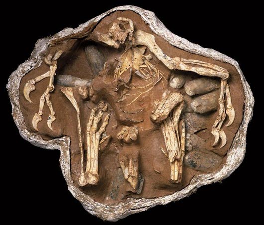
It is another theropod that takes centre stage this time, a dinosaur called Citipati osmolskae.  Citipati was a member of the Oviraptoridae, a very bird-like family of Late Cretaceous dinosaurs.   A number of C. osmolskae specimens are known, including individuals found preserved along with nests of their own eggs.  One such beautifully preserved specimen, MPC-D 100/979, nick-named “Big Mamma” was studied using scanning and transmission electron microscopy to identify minute details of both the fossil’s surface and its internal structure in a bid to assess whether any organic material persisted in the fossilised claws.

The Citipati osmolskae Specimen Studied “Big Mamma”

Citipati osmolskae fossil.

The Citipati fossil sitting on a nest “Big Mamma”.

Picture credit: M. Ellison/The American Museum of Natural History

The researchers have identified tell-tale signatures that suggest the preservation (on a microscopic level), of the original keratinous claw sheath that covered its digits.  This study adds to the growing body of evidence that original organic materials can be preserved over time.

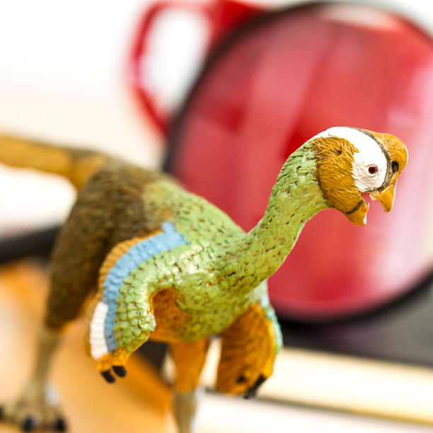
A Model of Citipati osmolskae (Safari Ltd)

Wild Safari Prehistoric World Citipati osmolskae dinosaur figure.

Citipati dinosaur model.

The Oviraptorid Citipati osmolskae

Citipati osmolskae was a theropod dinosaur that lived in what is now Mongolia during the Late Cretaceous.  In 1995, a particularly well-preserved specimen of Citipati was recovered from the Djadokhta Formation of Mongolia.  The specimen was found in a brooding position on a nest of eggs.  It is thought that the adult and nest were rapidly buried when a sand dune collapsed on top of them.

The nearly complete remains have been beautifully preserved and it had been noted that there was a thin lens of white material extending beyond one of the bony claws on a forelimb that differed in texture and colour from both the surrounding matrix and the bone.  It was also located where a claw sheath would be.   At around three metres in length (mostly tail), Citipati is one of the largest oviraptorids known, it is thought to be very closely related to Oviraptor.

For models and replicas of oviraptorids including Citipati (whist stocks last): Safari Ltd. Dinosaurs and Prehistoric Animal Figures.

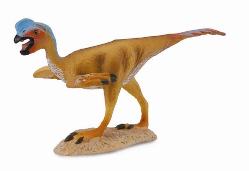
An Illustration of a Typical Oviraptorid

A model of an Oviraptor.

A typical oviraptorid – Oviraptor philoceratops.

Picture credit: Everything Dinosaur

Studying the Claws of “Big Mamma”

Alison Moyer, former PhD student at North Carolina State University, who is currently a postdoctoral researcher at Drexel University and lead author of a paper describing the research, wanted to find out if the material from Citipati was a claw sheath and if so, whether any original beta-keratin had preserved.

In extant Aves, (birds), claw sheaths cover the claw at the end of a digit much like fingernails in humans. The sheaths in modern birds are composed of two types of keratin, alpha-keratin, the softer form found on the interior of the sheath; and beta-keratin, a harder and more durable keratin that comprises the sheath’s exterior.  The team set out to discover whether they could detect the presence of beta-keratin.

Moyer and her university colleagues first used powerful scanning electron microscopy to assess the material.  The results showed that the sample was structurally similar to claw sheaths from modern birds, so the team decided to proceed with immunohistochemical (IHC) testing, this would identify the nature of the material preserved.

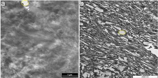
Highly Magnified Images Comparing the Internal Structure of Citipati Claw Sheath with the Internal Structure of an Ostrich Claw Sheath

Citipati dinosaur claw sheath studied.

TEM micrographs of (a) ostrich claw sheath and (b) Citipati claw sheath show similar structure.

Picture credit: Alison Moyer

The images above are highly magnified micrographs of (a) ostrich claw sheath and (b) Citipati claw sheath which show a similar structure.  In both samples, parallel fibres can be seen running diagonally, and identical voids (yellow outlines) were observed among the fibres in both samples.

Immunohistochemical Testing (IHC)

IHC testing utilises antibodies that react against a particular protein.  If the protein is present, the antibodies bind to small regions of the protein and indicate where the protein is located in the tissue.  Moyer used beta-keratin antibodies derived from modern bird feathers.  In initial IHC testing, results were inconclusive, which led Moyer to look more closely at the specimen.  She found an unusually high concentration of calcium in the fossil claw, much higher than would be found in claws from the living birds used in comparison (such as Ostrich claw material), or from the sediment surrounding the fossil.

The team postulated that the calcium might be affecting results.  The calcium was eliminated and further IHC testing on the claw sheath material was conducted.  After the calcium was removed, the antibodies reacted much more strongly, indicating the presence of beta-keratin and preservation of original molecules.

Dinosaur Claw Proteins

Moyer explained:

“It’s probable the incorporation of calcium in the tissue helped preserve it, but that same calcium had to be removed in order to see the underlying molecular composition.  Because this study used multiple, well-tested methods, it not only supports the longevity of proteins in the rock record, it reveals a lot about how these might be preserved.”

The researchers conclude that calcium chelation greatly increased antibody reactivity, suggesting a role for calcium in the preservation of this type of fossil material.

The scientific paper: Microscopic and Immunohistochemical Analyses of the Claw of the Nesting Dinosaur, Citipati osmolskae, Proceedings of the Royal Society B.

Everything Dinosaur acknowledges the help of North Carolina State University in the compilation of this article.

Mineralised Blood Vessels in a Hadrosaur: Duck-billed Dinosaur Blood.

Details of a Documentary about the work of Dr Schweitzer: The Search for Dinosaur DNA.

Visit the website of Everything Dinosaur: Everything Dinosaur.

13 11, 2016

Appreciating the Remarkable Dunkleosteus

By |2024-05-06T15:31:31+01:00November 13th, 2016|Categories: Dinosaur and Prehistoric Animal News Stories, Dinosaur Fans, Main Page, Photos/Pictures of Fossils|0 Comments

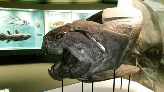
Dunkleosteus Exhibit at the Naturmuseum Senckenberg

At the Naturmuseum Senckenberg (Frankfurt, Germany), visitors are treated to a spectacular gallery featuring that most diverse group of vertebrates, the fish.   Fans of Palaeozoic fossils can see several specimens on display, including a beautiful Dunkleosteus exhibit.

The Anterior Portion of Dunkleosteus on Display

Dunkleosteus on display.

The spectacular Dunkleosteus exhibit.

Picture credit: Everything Dinosaur

Greeting you at the gallery entrance area is a cast of the plated skull and jaws of the giant Devonian predator Dunkleosteus.  It certainly is a most impressive sight.  Sharp-eyed readers can spot the John Sibbick illustration that can be seen behind the replica skull with its shear-like jaws.  In the background part of the fossil fish display can be seen.  It features a life-size model of a Coelacanth (Latimeria).

Standing Guard at the Fish Gallery Entrance – Dunkleosteus

A Dunkleosteus exhibit.

A Dunkleosteus cast on display.

Picture credit: Everything Dinosaur

A Powerful Placoderm

Dunkleosteus was a huge, prehistoric fish with an armoured head made up of several bony plates that covered over thirty percent of the entire animal’s length. Larger specimens had dermal armour that was up to five centimetres thick.  The placoderms (armoured fish); evolved in the Silurian period from ancestors that had no teeth, instead this group of fish developed a pair of extremely sharp bony plates that hung from the top jaw, the edges of the lower jaw were also bony and as sharp as a razor.  The jaws could be slammed together like a pair of self-sharpening shears.

For models and replicas of prehistoric fish and other extinct animals: Prehistoric Animal Figures from PNSO.

Dr John Newberry formally named and scientifically described this apex predator in 1873.  The genus name honours the famous American palaeontologist Dr David Dunkle of the Cleveland Museum of Natural History.

As for size, a number of estimates have been published.  The Everything Dinosaur fact sheet sent out with Dunkleosteus model purchases suggests a maximum length of about nine to ten metres, with a maximum body mass of around four tonnes.

The CollectA 1:20 Scale Replica of Dunkleosteus Compared to the Museum Specimen

The CollectA 1:20 scale Dunkleosteus compared to a museum specimen.

A comparison between the new for 2018 CollectA 1:20 scale replica of Dunkleosteus and a museum specimen.  Picture credit: Everything Dinosaur.

Picture credit: Everything Dinosaur

To view the CollectA Prehistoric Life scale model series: CollectA Deluxe Prehistoric Animal Figures and Replicas.

12 11, 2016

Apex Predator from Late Cretaceous Antarctica Described in New Research

By |2024-05-06T15:32:02+01:00November 12th, 2016|Categories: Dinosaur and Prehistoric Animal News Stories, Dinosaur Fans, Main Page|0 Comments

Largest Mosasaur from Antarctica Described – Kaikaifilu hervei

A team of international researchers writing in the journal “Cretaceous Research”, have published details of the largest mosasaur to be identified from Upper Cretaceous strata in Antarctica.  The newly described marine reptile has been named Kaikaifilu hervei, the scientists, which included Rodrigo Otero, a palaeontologist at the University of Chile and the lead researcher on the study, named the mosasaur after “Kai-Kai filú,” an almighty giant reptile that ruled the sea in legends from the Mapuche culture from southern Chile and Argentina.

Kaikaifilu hervei

The species name honours Francisco Hervé (University of Chile), who has done much to improve our understanding of the palaeoclimate of Chile and Antarctica.

Kaikaifilu hervei fossil material was found by a Chilean palaeontology expedition to the remote Seymour Island (sometimes called Marambio Island), off the Antarctic peninsula.  This location has attracted a number of scientific expeditions in recent years as the strata exposed on parts of the island bridges the time period from the end of the Cretaceous and into the Palaeocene Epoch. The fossils, which include a fragmentary skull, plus a partial left humerus, elements from the jaw and about thirty teeth have been dated to around 66 million-years ago (López de Bertodano Formation).

The Estimated Size of Kaikaifilu hervei

Kaikaifilu hervei size comparison

Kaikaifilu hervei estimated to be around ten metres in length.

Picture credit: Otero, R, A, Cretaceous Research with additional annotation by Everything Dinosaur

An Apex Predator

Based on better known mosasaurs, the scientists estimate that K. hervei reached lengths in excess of ten metres.  It was probably an apex predator preying on the plesiosaurs that shared its shallow, marine environment.  The fossil skull is only the second mosasaur skull known from Antarctica.  The other mosasaur skull found in Antarctica represents the closely related, but earlier Taniwhasaurus antarcticus, which is estimated to have been about half the size of K. hervei.  T. antarcticus fossils have been found on James Ross Island, part of the same archipelago as Seymour Island.

The Fossil Excavation Site on Seymour Island

Mosasaur excavation site Seymour Island.

The desolate excavation site (K. hervei) Seymour Island in Antarctica.

Picture credit: Otero, R, A, Cretaceous Research

For models and replicas of plesiosaurs and mosasaurs: Papo Dinosaur and Prehistoric Animal Figures.

A Hunter of Plesiosaurs

Lead author of the study, Rodrigo Otero stated:

“Prior to this research, the known mosasaur remains from Antarctica provided no evidence for the presence of very large predators like Kaikaifilu, in an environment where plesiosaurs were especially abundant.  The new find compliments one expected ecological element of the Antarctic ecosystem during the latest Cretaceous.”

Kaikaifilu has been classified as a member of the Tylosaurinae sub-family of the Mosasauridae, as such, it was closely related to the Tylosaurus genus known from the United States.  The thirty or so teeth show heterogeneity (different shapes), this suggests that species of mosasaur named from just their different shaped teeth may in fact represent the Kaikaifilu genus and the researchers have postulated that the discovery of heterogeneous teeth on Seymour Island may lead to some revision amongst the Mosasauridae.

The scientific paper: “Kaikaifilu hervei gen. et sp. nov., A New Large Mosasaur (Squamata, Mosasauridae) from the Upper Maastrichtian of Antarctica”.

The website of Everything Dinosaur: Everything Dinosaur.

10 11, 2016

“Stuck in the Mud” Dinosaur Provides Insight into Oviraptorosaur Radiation

By |2023-05-09T07:33:03+01:00November 10th, 2016|Categories: Dinosaur and Prehistoric Animal News Stories, Dinosaur Fans, Main Page|2 Comments

Tongtianlong limosus – One Very Unusual Fossil Posture

A team of international researchers, scientists from the Chinese Academy of Sciences, Dongyang Museum (Zhejiang Province, China) and Edinburgh University have announced the discovery of a new genus of bird-like dinosaur.  This newest member of the Oviraptoridae family, most likely met its demise when it became trapped in mud. The dinosaur has been named Tongtianlong limosus.

Tongtianlong limosus

The animal was preserved with its forelimbs splayed out and outstretched as if beseeching someone or something to help pull it to safety. This dinosaur’s run of bad luck did not end in the Maastrichtian, the specimen was nearly blown up when construction workers were preparing the ground for the building of a new high school.

Two Views of the Fossil Skeleton Material (Tongtianlong limosus)

Tongtianlong fossils.

Two views of the holotype fossil material of Tongtianlong (a) dorsal view and (b) lateral view. Scale bar = 10 cm.

Picture credit: Nature Scientific Reports (Junchang Lü et al)

Late Cretaceous Southern China was Oviraptorosaur Country

The fossil material, a near complete, three-dimensional specimen was saved by the construction workers, those who had narrowly avoided blasting the last resting place of this sheep-sized dinosaur to smithereens, a drill hole for an explosive charge can even be seen in the pelvic area of the holotype specimen (see photograph above).   The builders did their best to retrieve as much of the fossil material as they could.  With such an amazing skeleton to study, the researchers were quickly able to identify it is an oviraptorid and several unique characteristics led them to describe it as a new species.

An Illustration of Tongtianlong limosus Trapped in Mud

The feathered dinosaur Tongtianlong limosus mired in mud.

Tongtianlong trapped in mud.

Picture credit: Zhao Chuang

A Sixth Species of Oviraptorosaur

This is the sixth species of oviraptorosaur* to have been discovered in the Upper Cretaceous strata (Nanxiong Formation), exposed in Ganzhou Province (south-eastern China).  The prevalence of oviraptorid fossil material may represent some form of preservation bias, but more likely, the fossil material indicates that this part of world was home to a variety of different types of oviraptorosaur.

Analysis of skull shape and the jaws suggest that each type of oviraptorosaur had evolved to exploit different food resources.  The number of different kinds of this type of dinosaur found in this locality suggests that oviraptorids were undergoing a radiation and rapidly evolving.  As such, these feathered, bird-like dinosaurs, may have been one of the last groups of dinosaur to undergo evolutionary radiation before their extinction.

The Skull of Tongtianlong limosus with Line Drawing  Below

Skull and line drawing of Tongtianlong limosus.

The skull of Tongtianlong with an accompanying line drawing.  Scale bar = 5 cm.

Picture credit: Nature Scientific Reports (Junchang Lü et al)

Key

aof = antorbital fenestra, bc = braincase, d =dentary, emf = external mandibular fenestra, eo = exoccipital, f -= frontal, j = jugal, l = lacrimal, ltf = lower temporal fenestra, m = maxilla, n = nasal, nar = narial opening, npc = nasopharyngeal canal, o = orbit (eye socket see note), p = parietal, pm = premaxilla, pno = pneumatic opening, po = postorbital, q = quadrate, qj = quadratojugal, sq = squamosal and stf = supratemporal fenestra

Lots of Different Oviraptorid Dinosaurs in the Ecosystem

Tongtianlong is the sixth oviraptorosaurian taxon named from the Nanxiong Formation in this part of south-eastern China.  All these taxa have been named since 2010, the genera are:

  • Banji (B. long) named in 2010 – size unknown.
  • Ganzhousaurus (G.  nankangensis) named in 2013 – size not known.
  • Jiangxiasaurus (J. ganzhouensis) named in 2013 – size not known
  • Nankangia (N. jiangxiensis) named in 2013 – 2.5 metres long.
  • Huanansaurus (H. ganzhouensis) named in 2015 – 2.5 metres in length.

For models and replicas of Asian oviraptorid dinosaurs and other prehistoric animals: Age of Dinosaurs Figures from PNSO.

In the scientific paper the researchers explore the possible reasons why there were so many oviraptorids in Late Cretaceous southern China.  It could be due to “taxonomic inflation”, some of the specimens described could in fact be males or females of already known species or perhaps the fossils could represent already described species of dinosaur but different growth stages.

The scenario the scientists favour is that the fossils are documentary evidence supporting the idea that there was a genuine radiation of oviraptorosaurs.

The Scientific Paper: “A Late Cretaceous Diversification of Asian Oviraptorid Dinosaurs: Evidence from a New Species Preserved in an Unusual Posture”.

Visit Everything Dinosaur’s website: Everything Dinosaur.
8 11, 2016

Rapid Recovery of Patagonian Plant-Insect Associations Reported in New Scientific Paper

By |2024-05-06T15:32:40+01:00November 8th, 2016|Categories: Dinosaur and Prehistoric Animal News Stories, Dinosaur Fans, Main Page, Palaeontological articles|0 Comments

Research Suggests Southern Hemisphere a Relatively Safe Haven after End Cretaceous Impact Event

Life in much of the Southern Hemisphere seems to have recovered more quickly than ecosystems further north after the catastrophic Yucatan Peninsula impact event that marked the end of the Cretaceous.

Furthermore, the southern parts of our planet may have provided a biodiversity refuge after the Cretaceous/Palaeogene mass extinction.  These are the conclusions drawn in a newly published scientific paper written by researchers based in the United States and Argentina.

Fossil Leaves and Insect Damage

The scientists, which included graduate student Michael Donovan and his supervisor, Professor Peter Wilf (Department of Geosciences, Pennsylvania State University), examined thousands of fossil leaves preserved in strata that represent the latest stage of the Cretaceous (Maastrichtian) and the earliest part of the Palaeocene (Danian).  The fossils were examined under high magnification to identify traces of insect-feeding damage.  From this data, the team were able to calculate how robust and diverse the ecosystem was at each point in deep time represented by the different stratigraphical layers.

An Analysis of Insect Feeding Damage on Fossil Leaves

Leaf miner damage in a Cretaceous leaf.

Mining in a leaf. Initial serpentine phase packed with frass followed by a dramatic widening into a
blotch phase on leaf and b, close-up
of mine path in a.

Picture credit: Pennsylvania State University

Insect Damage on Leaves – A Measure of the Health of Terrestrial Food Chains

Biologists and botanists have used the number of plant/insect associations recorded in modern-day terrestrial ecosystems to assess the biodiversity of the food chain.  These same principles can be applied to a study of fossil biota and as such, a study that looks at plant-insect associations across the K/Pg boundary can provide palaeontologists with a more complete understanding of how ecosystems coped and bounced back from a mass extinction event.

In an analysis of 3,646 fossil leaves from the latest Maastrichtian as well as from the Danian faunal stages preserved in strata located in Chubut Province, Patagonia (southern Argentina), the team concluded that it took approximately four million years for plant-insect associations to recover after the extraterrestrial impact event.  Studies of insect-plant associations in strata of similar age but much closer to ground zero (western North America), indicate that it took at least nine million years for these associations to recover.

Insect Damage on Fossil Leaves from the Palaeocene

Fossil leaves showing insect damage.

Insect damage from fossil leaves (Danian faunal stage of the Palaeocene).

Picture credit: Pennsylvania State University

The picture above shows examples of Palaeocene fossil leaves with insect feeding damage.  In photograph (k), skeletonised leaf tissue as a result of insect feeding is shown and (l) shows a portion of the leaf margin of Dryophyllum australis with feeding evidence along the margin.   This new paper, published in the journal “Nature Ecology and Evolution” supports the emerging hypothesis that the impact of the extraterrestrial impact event on ecosystems was variable, particularly in the Southern Hemisphere.

Ecosystems in the Southern Hemisphere Recovered Faster

In general terms, in areas closer to the impact site, such as in western North America, little evidence has been found to support the idea that many different types of insect survived.  However, ecosystems seem to have recovered much faster in South America.  Studies of microscopic plankton and pollen have also provided evidence that life bounced back more quickly in the Southern Hemisphere than in the north.  The palaeolatitude of the Chubut Province was around 50 degrees south, such locations may have provided a refuge for biota that could then migrate northwards as conditions improved.

Intriguingly, not all fossil sites in the Southern Hemisphere show signs of biota refuge status at the K/Pg boundary.  In June of this year, Everything Dinosaur published an article on a study of the Upper Cretaceous rocks and Lower Palaeocene strata exposed on Seymour Island in the Antarctic.  This research indicated that there was a dramatic and rapid extinction event in southernmost marine environments.

To read more about this study: Global Catastrophe Caused End Cretaceous Mass Extinction.

A spokesperson from Everything Dinosaur remarked that this new study could have important implications.  Further research is needed to ascertain the global recovery rate.  However, this study provided compelling evidence to indicate that some parts of the world recovered more quickly than others.

Visit Everything Dinosaur’s website: Everything Dinosaur.

28 10, 2016

A Rare Dinosaur Brain from Bexhill-on-Sea

By |2024-05-06T15:16:18+01:00October 28th, 2016|Categories: Dinosaur and Prehistoric Animal News Stories, Dinosaur Fans, Main Page|0 Comments

Fossilised Dinosaur Brain Tissue Identified for the First Time

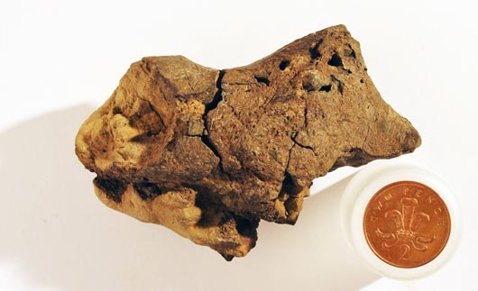
Bexhill-on-Sea may not be synonymous with ground-breaking palaeontology, but a visit to the local museum will reveal that dinosaur bones and teeth have been found in this part of East Sussex (southern England).  However, a small fossil measuring a little over ten centimetres long and five centimetres across, a fossil found on the foreshore area of the beach by amateur fossil collector Jamie Hiscocks in 2004, has proved to be the first fossilised dinosaur brain known to science.

A View of the Fossil – Preserved Parts of the Exterior of a Dinosaur’s Brain

The fossilised brain of a dinosaur.

A small pebble from the foreshore at Bexhill-on-Sea is actually part of the fossilised brain of a dinosaur.

Picture credit: Jamie Hiscocks/Press Association

The Brain from an Iguanodontid

Although the genus cannot be identified, the researchers which include Dr David Norman and Dr Alex Liu (Cambridge University), are confident that this 133-million-year-old (Lower Cretaceous) fossil, came from a large ornithopod, an iguanodontid.  Fossils of these herbivorous dinosaurs are known from the strata that is exposed in this area and the brain displays distinct similarities to modern-day archosaurs, the closest living relatives to the Dinosauria, crocodiles and birds.

For models and replicas of iguanodontids and other dinosaurs: CollectA Age of Dinosaurs Popular Figures and Models.

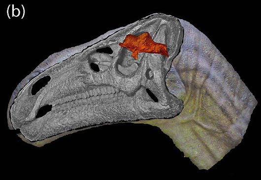
The Fossil Placed in the Correct Anatomical Position Within the Skull of an Iguanodontid

Showing the location of the dinosaur brain element.

The location of the fossil within the skull of the iguanodontid.

Picture credit: Oxford University and Cambridge University

Meningeal Blood Vessels Identified

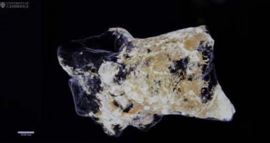
Scanning microscopy undertaken by colleagues at the University of Western Australia and three-dimensional animations provided by the University of Manchester reveal the meninges, the tough tissues surrounding the actual brain, as well as tiny capillaries and portions of the adjacent soft tissues representing the cortex.  The researchers describe these details as “mineralised ghosts”.

An Image from the Three-Dimensional Animation Provided by the University of Manchester

A computerised, three-dimensional image of the brain.

A still from the Manchester University animation which shows the fine detail on the fossil’s exterior.

Picture credit: University of Cambridge/University of Manchester

This remarkable research, a first for palaeontologists who specialise in studying the Dinosauria, has been published in a Special Publication of the Geological Society of London.  The paper has been published in tribute to Professor Martin Brasier (Oxford University), who sadly died in 2014 following a road traffic accident.  Professor Brasier had coordinated the research alongside Dr David Norman.  One of the co-authors of the paper Dr Alex Liu was a PhD student of Professor Brasier at the time the research project was started.

Dr Liu, explained the importance of this study:

“The chances of preserving brain tissue are incredibly small, so the discovery of this specimen is astonishing.”

A Dead Dinosaur with its Head Stuck Upside Down in Stagnant Water

According to the researchers, the reason this particular piece of brain tissue has been so well-preserved is that the dinosaur’s brain was essentially ‘pickled’ in a highly acidic and low-oxygen body of water.  Shortly after this dinosaur died, the corpse ended up in a shallow, stagnant pool, perhaps a bog or part of a swamp.  The skull ended up buried in the fine sediment at the bottom and this allowed the soft tissue structures to be mineralised before they began to decay.  This permitted the high degree of detail to be preserved.

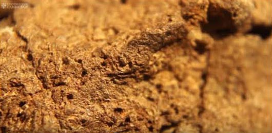
A Magnified View of the Exterior of the Specimen

A closer view of the dinosaur brain fossil.

A close view of the fine structures preserved on the fossil material.

Picture credit: Oxford University and Cambridge University

Dr Norman commented:

“What we think happened is that this particular dinosaur died in or near a body of water, and its head ended up partially buried in the sediment at the bottom.  Since the water had little oxygen and was very acidic, the soft tissues of the brain were likely preserved and cast before the rest of its body was buried in the sediment.”

In collaboration with researchers from the University of Western Australia, the scientists employed scanning electron microscopy in order to identify the tough membranes, or meninges, that surrounded the brain itself, as well as strands of collagen and blood vessels.

A Highly Magnified Portion of the Brain Showing the Meningeal Blood Vessels

Meningeal blood vessels from the dinosaur brain.

Tubular structures identified on the exterior of the brain interpreted as representing meningeal blood vessels.

Picture credit:  Oxford University and Cambridge University

Bird Brains and the Brains of Crocodilians

Such is the exceptional preservation of the specimen that the scientists have been able to identify mineralised structures that represent tissues from the cortex (the outer layer of neural tissue of the brain), along with delicate, tiny capillaries.  These preserved tissues, especially the meninges are homologous to the brain tissues of extant Aves and crocodiles, close relatives of dinosaurs.

Scanning Electron Microscopy Reveals Remarkable Details

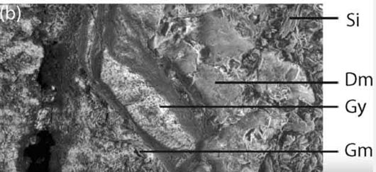
Scanning electron microscope images of the dinosaur brain.

The scanning electron microscope images revealed details of the internal structure of the outermost surface of the brain.

Picture credit:  Oxford University and Cambridge University

The picture above shows a highly magnified section of the fossil, sinuses as well as the tough and fibrous tissues protecting the actual brain can be made out.  Could “Dm” represent the Dura mater, the tough membrane that helps to protect the brain?

A Typical Reptilian Brain

Reptile brains (and bird brains for that matter), are shaped very differently from our own brain with its substantial cerebellum.  In most reptile brains, the brain is sausage-shaped, surrounded by a dense region of blood vessels and thin-walled vascular chambers (sinuses), the provide a blood drainage system.  The reptile brain only occupies about fifty percent of the actual cranial cavity.

In contrast, the tissue in the fossilised brain appears to have been pressed directly against the skull, raising the possibility that some dinosaurs had large brains, with a greater volume which filled much more of the cranial cavity.  With this one specimen to study, the researchers urge caution against drawing too many conclusions as to the intelligence of dinosaurs.  It is likely that post-mortem, the brain got pressed against the bony roof of the cranial cavity.  It is difficult to infer brain size based on the data available.

Not Sure How Big the Brain Was

Dr Norman added:

“As we can’t see the lobes of the brain itself, we can’t say for sure how big this dinosaur’s brain was.  Of course, it’s entirely possible that dinosaurs had bigger brains than we give them credit for, but we can’t tell from this specimen alone. What’s truly remarkable is that conditions were just right in order to allow preservation of the brain tissue, hopefully this is the first of many such discoveries.”

A Dinosaur Brain

Fossil hunter Jamie Hiscocks, the finder of the specimen back in 2004 said:

“I have always believed I had something special.  I noticed there was something odd about the preservation, and soft tissue preservation did go through my mind.  Martin realised its potential significance right at the beginning, but it wasn’t until years later that its true significance came to be realised.”

Everything Dinosaur acknowledges the help of Cambridge University in the compilation of this article.

The scientific paper:

Martin D. Brasier et al.’ “Remarkable Preservation of Brain Tissues in an Early Cretaceous Iguanodontian Dinosaur.” Earth System Evolution and Early Life: A Celebration of the Work of Martin Brasier. Geological Society, London, Special Publications.

Visit Everything Dinosaur’s website: Everything Dinosaur.
Go to Top