All about dinosaurs, fossils and prehistoric animals by Everything Dinosaur team members.

Pictures of fossils, fossil hunting trips, fossil sites and photographs relating to fossil hunting and fossil finds.

16 04, 2022

A Juvenile Diamantinasaurus – Australia’s Smallest Sauropod Found to Date

By |2023-07-04T07:21:38+01:00April 16th, 2022|Categories: Adobe CS5, Dinosaur and Prehistoric Animal News Stories, Dinosaur Fans, Main Page, Palaeontological articles, Photos/Pictures of Fossils|0 Comments

A scientific paper has just been published describing the fossilised remains of a juvenile titanosaur from the Winton Formation of Queensland, Australia. The specimen has been assigned to the Diamantinasaurus taxon (D. matildae) and it represents the smallest sauropod described from fossils found in Australia to date.

Reconstructed skeleton of the juvenile Diamantinasaurus (D. matildae) compared to a human skeleton.
A model of the reconstructed skeleton of the juvenile Diamantinasaurus (D. matildae) compared to a human skeleton. The young dinosaur is estimated to have weighed around 4.2 Tonnes. Picture credit: Australian Age of Dinosaurs Museum.

About Ten Percent of the Skeleton Recovered

The fossils were discovered on Elderslie Station land which lies some 35 miles northwest of the town of Winton (Queensland). Landowners noticed fragments of a femur and dorsal ribs exposed on the surface (2012). Staff from the Australian Age of Dinosaurs Museum along with volunteers excavated the site and found the remainder of the fossil material representing about 10% of the total skeleton about a metre below the surface.

The postcranial material consists of cervical ribs, three incomplete dorsal vertebrae, sacral vertebrae and limb bones.

Views of the juvenile Diamantinasaurus fossils (AODF 663) a right humerus and right manual ungual with accompanying digital models.
Views of the juvenile Diamantinasaurus fossils (AODF 663) a right humerus and right manual ungual with accompanying digital models. Right humerus photographs in A, dorsal, B, anterior, C, ventral, D, medial, E, posterior, F, lateral views. Right humerus digital models in G, dorsal, H, anterior, I, ventral, J, medial, K, posterior, L, lateral views. The right manual ungual in M, proximal, N, dorsal, O, anterior, P, ventral, Q, posterior views. Right manual ungual digital models in R, proximal, S, dorsal, T, anterior, U, ventral, and V, posterior views. Note scale bar for humerus equals 10 cm and for the manual ungual 5 cm. Picture credit: Rigby et al.

A Young Titanosaur from the Late Cretaceous

Although age estimates for the Winton Formation vary, it has been informally divided into lower and upper members, with the Diamantinasaurus material coming from the “upper” portion which is regarded as Cenomanian to potentially the lowermost Turonian stages of the Late Cretaceous (approximately 95-89 million years ago).

The study of the juvenile titanosaur was led by Museum Research Associate Samantha Rigby who is undertaking a Master of Science (Research) at Swinburne University of Technology (Victoria, Australia), under the supervision of Dr Stephen Poropat who was one of the co-authors of the scientific paper published in the Journal of Vertebrate Palaeontology. Each bone from the specimen was scanned to create three-dimensional models to digitally compare them with other sauropod remains.

This comparison suggests the small specimen belongs to the Diamantinasaurus taxon though with juvenile characteristics, vertebrae which are unfused, minimal muscle scarring on the bones, smooth bone texture and marked proportional bone size differences when compared to adult titanosaur material.

Diamantinasaurus dorsal vertebrae and digital models.
Diamantinasaurus juvenile dorsal vertebrae with digital models. Dorsal vertebra photographs in A, dorsal, B, right lateral, C, anterior, D, left lateral, E, posterior views. Dorsal vertebra digital models in F, dorsal, G, right lateral, H, anterior, I, left lateral, J, posterior views. Note scale bar equals 10 cm. Picture credit: Rigby et al.

Allometric Growth

The fossil specimen (AODF 663) nicknamed “Oliver” is only the third specimen to be referred to the taxon Diamantinasaurus matildae. D. matildae was formally named and described in 2009: A Trio of New Dinosaurs from Down Under. The research team found that the bones of this small titanosaur grew allometrically, meaning that its bones changed shape and different parts of its body grew at different rates.

The limb bones are also narrower in width when compared to other Diamantinasaurus limb bones from older individuals. This suggests that as this titanosaur grew its limb bones became thicker and more robust to help support its enormous bulk.

A Juvenile Diamantinasaurus

Fossils of juvenile titanosaurs are rare and it is hoped that “Oliver” will provide important insights into the ontogeny of titanosaurs.

Everything Dinosaur acknowledges the assistance of a media release from the Australian Age of Dinosaurs Museum in the compilation of this article.

The scientific paper: “A juvenile Diamantinasaurus matildae (Dinosauria: Titanosauria) from the Upper Cretaceous Winton Formation of Queensland, Australia, with implications for sauropod ontogeny” by Samantha L. Rigby, Stephen F. Poropat, Philip D. Mannion, Adele H. Pentland, Trish Sloan, Steven J. Rumbold, Carlin B. Webster and David A. Elliott published in the Journal of Vertebrate Paleontology.

Visit the Everything Dinosaur website: Everything Dinosaur.

15 04, 2022

Juvenile Gorgosaurus Skulls Shed Light on Tyrannosaurid Growth Patterns

By |2024-12-29T22:41:08+00:00April 15th, 2022|Categories: Adobe CS5, Dinosaur and Prehistoric Animal News Stories, Dinosaur Fans, Main Page, Palaeontological articles, Photos/Pictures of Fossils|1 Comment

Researchers have described two beautifully preserved skulls of juvenile Gorgosaurus dinosaurs (G. libratus). The articulated specimens have enabled the scientists to build up a comprehensive picture of how these tyrannosaurids changed as they grew. Gorgosaurus and the much bigger, later tyrannosaurid T. rex exhibit similar changes in their skulls as they grow. This study will help palaeontologists to decipher tyrannosaur material and to determine the identity of previously misidentified specimens. It should also provide more evidence to help resolve the Nanotyrannus/T. rex debate.

Lateral views of juvenile Gorgosaurus skulls.

Skulls of the two new juvenile Gorgosaurus libratus specimens in lateral view. A, TMP 2009.12.14; B, TMP 2016.14.1. Note scale bar equals 10 cm. Picture credit: Voris et al.

Picture credit: Voris et al

Gorgosaurus libratus

Named and described in 1914 (Lambe), Gorgosaurus is known from dozens of fossil specimens found in the upper Campanian Dinosaur Park Formation of Alberta and Judith River Formation of Montana. It is one of the best sampled and researched of all the Late Cretaceous tyrannosaurs, but juvenile material is rare. The recent discovery of additional juvenile Gorgosaurus libratus specimens from the Dinosaur Park Formation, including two well-preserved skeletons with articulated skulls, provided researchers which include Jared Voris and Darla Zelenitsky (University of Calgary), along with collaborators from the University of Ohio, the University of Alberta and the Royal Tyrrell Museum, an opportunity to develop a map outlining how this dinosaur changed as it grew and matured.

Juvenile and adult Gorgosaurus skulls compared.

Illustrations of juvenile (left) and adult (right) skulls of Gorgosaurus in lateral (top) and dorsal views (bottom). Arrows and numbers indicate ontogenetically invariant autapomorphies of Albertosaurinae and Gorgosaurus as per emended diagnosis. Juvenile illustration based on TMP 2016.14.1 (lateral) and TMP 2009.12.14 (dorsal), adult illustration based on UALVP 10. Picture credit: Voris et al.

Picture credit: Voris et al

Sorting out Daspletosaurus Specimens

The research team, which also included Professor Phil Currie (University of Alberta), found that although the skulls of tyrannosaurs changed dramatically as they grew, several taxonomically informative traits remain present regardless of the age of the animal. This means that palaeontologists can use this information to determine which taxon is represented by juvenile fossil material.

Thanks to this research, two specimens previously identified as examples of immature Daspletosaurus individuals (coeval with Gorgosaurus) are instead confirmed as Gorgosaurus.

Gorgosaurus dentaries compared

Left dentaries in lateral view of A, small juvenile (TMP 1994.12.155); B, juvenile (TMP 2016.14.1); C, subadult (TMP 1991.36.500); D, young adult (ROM 1247); and E, adult (TMP 1967.9.164) specimens of Gorgosaurus libratus. Note the development of the autapomorphic dorsoventral expansion in the posterior region of the bone through ontogeny. Scale bar equals 10 cm. Picture credit: Voris et al.

Picture credit: Voris et al

Comparisons with Tyrannosaurus rex

The team also found that both Gorgosaurus and T. rex underwent similar anatomical changes over their lifespans, but at different times. The changes started later in Tyrannosaurus rex and occurred over a longer time interval, resulting in a larger size and longer lifespan for T. rex when compared to Gorgosaurus.

Comparing the growth stages of Gorgosaurus libratus and T. rex.

Comparison of the growth series of Gorgosaurus libratus (top) and Tyrannosaurus rex (bottom), demonstrating similar ontogenetic stages (and morphologies) occurring at similar relative size (percent of largest specimen skull length) but different body sizes and biological ages. Picture credit: Voris et al.

Picture credit: Voris et al

Implications for Nanotyrannus

Having identified a series of anatomical traits that can be relied upon to permit palaeontologists to confidently assign juvenile tyrannosaur skull fossils to a specific taxon, this allows some specimens considered small or “dwarf” forms such as Nanotyrannus (N. lancensis) to be revisited. Some of these specimens may have been misidentified, since key characteristics may not have developed in young individuals before death, but this new data set would allow closer scrutiny of the fossil material.

PNSO Nanotyrannus dinosaur model.

The new for 2021 PNSO Nanotyrannus dinosaur model.

The picture (above) shows a model of a Nanotyrannus by PNSO.

To view the range of PNSO prehistoric animal figures in stock: PNSO Prehistoric Animal Models.

The scientific paper: “Two exceptionally preserved juvenile specimens of Gorgosaurus libratus (Tyrannosauridae, Albertosaurinae) provide new insight into the timing of ontogenetic changes in tyrannosaurids” by Jared T. Voris, Darla K. Zelenitsky, François Therrien, Ryan C. Ridgely, Philip J. Currie and Lawrence M. Witmer published in the Journal of Vertebrate Paleontology.

Visit the award-winning Everything Dinosaur website: Dinosaur Toys.

14 04, 2022

An Important Update – Assessment Reports “Minor” Damage to Mill Canyon Track Site

By |2024-12-29T18:33:31+00:00April 14th, 2022|Categories: Adobe CS5, Dinosaur and Prehistoric Animal News Stories, Dinosaur Fans, Main Page, Palaeontological articles, Photos/Pictures of Fossils|0 Comments

An assessment of the Mill Canyon dinosaur track site north of Moab in Grand County, Utah by Bureau of Land Management regional palaeontologist Brent Breithaupt confirms that damage was caused to the Early Cretaceous tracks and trace fossils by a construction crew, but the damage is described as “minor”.

Dinosaur trackway.

Trace fossils (dinosaur footprints) preserved at Moab (Utah). Picture credit: Bureau of Land Management.

Picture credit: Bureau of Land Management

Essential Repairs and Maintenance at an Important Fossil Site

The site, which covers approximately 2.3 acres contains over 200 tracks and other trace fossils recording activity around a body of water at an Early Cretaceous lake (approximately 112 million years ago). A construction crew had been employed to undertake repairs and improvements to the site including the replacement of boardwalks. Members of the public became aware of the maintenance work and reported possible damage to the fossils caused by the construction crew.

Everything Dinosaur published a blog article on reports of the damage caused by Bureau of Land Management contractors: Dinosaur Tracksite Damaged and having had concerns raised about damage to the site, it was decided to conduct an assessment of the area in order to find the best way to protect the fossils whilst still permitting public access.

This Damage to the Track Site Should Not Have Occurred

The assessment concluded that the overall damage was minor. Even so, Brent Breithaupt wrote in the report that:

“This damage should not have occurred”.

The regional palaeontologist added, that if the project had not been stopped:

“It is likely that much greater damage would have occurred with increased construction activities”.

As the Bureau of Land Management failed to consult palaeontologists on the maintenance plans, crews did not know which areas of the site to avoid. The incident was described in the report as “unfortunate” and the damage “could have been avoided”.

Mill Canyon tracksite.

At least six different dinosaur tracks have been deciphered at Moab (Utah). Picture credit: Bureau of Land Management.

After the report was released, the Bureau of Land Management has confirmed that an additional environmental assessment would be undertaken, the public would be consulted and palaeontologists involved in future work at the location to supervise activities. The Bureau of Land Management reported that it “remains committed to protecting plant and animal fossils on our public lands”.

The Everything Dinosaur website: Dinosaur Toys.

12 04, 2022

Ichthyosaurs Had Blubber According to a New Study

By |2024-12-29T18:06:04+00:00April 12th, 2022|Categories: Adobe CS5, Dinosaur and Prehistoric Animal News Stories, Dinosaur Fans, Main Page, Palaeontological articles, Photos/Pictures of Fossils|0 Comments

Researchers from the Smithsonian Institute (Washington), the University of Oslo and the Ludwig Maximilian Universität (Munich), have published a paper that describes two ichthyosaur specimens from the famous Upper Jurassic Solnhofen deposits of southern Germany. The fossils, an almost complete Aegirosaurus (JME-SOS-08369) and a tail (JME-SOS183), reveal extensive soft tissue preservation, analysis of which indicates the presence of blubber in these marine reptiles.

Late Jurassic Aegirosaurus sp. (Solnhofen)

Late Jurassic Aegirosaurus sp. (JME-SOS-08369) shown in (A) in normal light and (B) composite picture in UV light indicating location of further close-up images included in the scientific paper. Interpretative drawing (C). Note scale bar equals 20 cm. Picture credit: Delsett et al.

Picture credit: Delsett et al

These amazing fossil specimens representing marine reptiles that lived around 150 million years ago will help scientists to better understand how soft tissue can be preserved in the carcases of vertebrates deposited on the seafloor.

For models and replicas of ichthyosaurs and other prehistoric animals: Ichthyosaurs and Prehistoric Animal Models (CollectA).

Aegirosaurus

Aegirosaurus is a genus of ichthyosaur within the Ophthalmosauridae family. Its fossils are associated with the Upper Jurassic limestone deposits of Solnhofen in southern Germany. These deposits are famous for their vertebrate fossils, although ichthyosaur material is rare. Fossils ascribed to this genus have also been found in Lower Cretaceous strata in south-eastern France (Fischer et al, 2011). This discovery suggests that most Late Jurassic ichthyosaurs came through the end Jurassic extinction and continued to thrive in the Early Cretaceous.

Aegirosaurus was an active, nektonic pursuit predator, probably feeding on small fish and squid.

Ophthalomosaurid model

A replica of Ophthalomosaurus, an ichthyosaur similar in body shape to Aegirosaurus.  Picture credit: DPA.

Picture credit: DPA

Evidence for Blubber

The nearly complete ichthyosaur skeleton (JME-SOS-08369) was excavated in 2009, whereas the second specimen involved in this study (the tail), was originally found in 1926, but not formally described. No genus has been assigned to the tail specimen, although the researchers confidently assign it to the Ophthalmosauridae.

Ichthyosaur tail fossil.

The Late Jurassic ichthyosaur tail (specimen number JME-SOS2183) shown in (A) regular light, under ultraviolet light (B) with an interpretative line drawing (C). Note scale bar equals 10 cm. Picture credit: Delsett et al.

Picture credit: Delsett et al

Soft tissue samples were analysed using X-ray diffraction (XRD) and scanning electron microscopy (SEM) coupled with energy dispersive spectroscopy (EDS) analysis. The analyses confirm the presence of the phosphate mineral apatite, with phosphate most likely derived from the body itself. In addition, a yellow-coloured, amorphous substance was identified, the researchers postulate that this substance represents decomposed blubber.

Aegirosaurus fossil skull with interpretative line drawing.

Skull from Late Jurassic Aegirosaurus sp. (JME-SOS-08369) with (A) interpretative drawing and (B) photograph. Note scale bar equals 5 cm. Picture credit: Delsett et al.

Picture credit: Delsett et al

Identifying Adipocere

The researchers conclude that the detailed analysis of the yellowish, amorphous substance indicates that it is adipocere. This is late-stage post-mortem decomposing fatty acids produced by microorganisms under low oxygen conditions. As adipocere is a typical breakdown product of animal blubber, it is postulated that these ichthyosaurs had blubber to help insulate them, just as many extant marine mammals do. The paper does not address any endothermic implications for this conclusion.

Understanding Ichthyosaur Taphonomy

The two ichthyosaur specimens with their extensive soft tissue preservation will help scientists to interpret the taphonomy (how fossils are preserved) of Solnhofen Archipelago vertebrates. Future research will focus on microscopical and geochemical analysis of different parts of the specimens that have the potential to reveal more information about tissue types.

In addition, the beautifully preserved fossils hold the potential for investigations into the locomotion of ophthalmosaurids, helping scientists to better understand how these marine reptiles moved through the water.

The scientific paper: “The soft tissue and skeletal anatomy of two Late Jurassic ichthyosaur specimens from the Solnhofen archipelago” by Lene L. Delsett​, Henrik Friis, Martina Kölbl-Ebert and Jørn H. Hurum published in PeerJ.

The Everything Dinosaur website: Prehistoric Animal Figures.

7 04, 2022

Evidence of Triceratops Fighting Rivals

By |2024-12-29T17:26:42+00:00April 7th, 2022|Categories: Adobe CS5, Dinosaur and Prehistoric Animal News Stories, Dinosaur Fans, Main Page, Photos/Pictures of Fossils|0 Comments

Scientists have analysed a hole in the fossil skull of a large Triceratops and concluded that the injury was caused by another Triceratops. This study suggests that Triceratops engaged in fights with other members of their species (intraspecific combat).

A specimen of Triceratops (T. horridus) referred to as Big John was discovered in 2014 in the Upper Cretaceous Hell Creek Formation (Montana, USA). There is a hole (fenestra), in the right squamosal. The neck shield is perforated and researchers from the University of Chieti-Pescara, the University of Bologna in collaboration with other research institutes conducted detailed tests on the fossilised bone surrounding this perforation.

Evidence of intraspecific combat in Triceratops.

The specimen of Triceratops horridus known as “big John” suggest that an injury to the neck frill was caused by intraspecific combat. Picture credit: Ferrara A., and Briano I.

Chemical Analysis

Extracranial fenestrae in ceratopsian neck frills had been interpreted as evidence of injuries that resulted from intraspecific combat. To evaluate this hypothesis the researchers conducted extensive tests on the fossil bone immediately surrounding the hole in the neck frill. Microscopy analysis revealed newly formed and healing bone, with histological signs typical of the bone remodelling phase associated with recovery from an injury. In addition, chemical analysis revealed typical signatures associated with bone re-growth and healing.

Triceratops Fighting

The picture (below) shows two Triceratops dinosaurs fighting. These are two models from the Eofauna Scientific Research model range. The models are called “Dominant” and “Cryptic”.

To view the Eofauna Scientific Research range: Eofauna Scientific Research Prehistoric Animal Models.

Eofauna Scientific Research Triceratops dinosaur models.

The Eofauna Scientific Research 1:35 scale Triceratops models do battle (Cryptic and Dominant).

The researchers conclude that histological and microanalytical analyses indicate that the squamosal fenestra of Big John is the result of a traumatic event, which might indeed have occurred during a fight with another Triceratops.

The scientific paper: “Histological and chemical diagnosis of a combat lesion in Triceratops” by Ruggero D’Anastasio, Jacopo Cilli, Flavio Bacchia, Federico Fanti, Giacomo Gobbo and Luigi Capasso published in Scientific Reports.

Visit the Everything Dinosaur website: Everything Dinosaur.

6 04, 2022

A New, Large Theropod Trackway from Spain is Described

By |2024-12-29T17:22:56+00:00April 6th, 2022|Categories: Adobe CS5, Dinosaur and Prehistoric Animal News Stories, Dinosaur Fans, Main Page, Palaeontological articles, Photos/Pictures of Fossils|0 Comments

Scientists have identified a trackway made by a large theropod dinosaur as it crossed shallow water, the tracks are exceptionally wide and reveal that this unfortunate predator probably had a dislocated toe.

Writing in the open access journal PLOS One, the researchers describe an exceptionally wide trackway (LH-Mg-10-16), made by a meat-eating dinosaur at a site at the Las Hoyas locality (La Huérguina Formation), in central Spain. The trackway consists of six prints (three left and three right prints) and has been interpreted as representing a large theropod dinosaur crossing shallow water, blanketed by a microbial mat.

Views of the LH-Mg-10-16 theropod trackway.

Views of the LH-Mg-10-16 theropod trackway. Cartography done in the field (A), (B) scanned surface of the trackway, mounted from a set of colour photos. Colour ramp, showing the depths and the countours of the site (in cm). Note scale bar equals 1 metre. The cartography is reprinted from Las Hoyas a Cretaceous wetland, Gibert JM et al. under a Creative Commons BY licence, with permission from Dr. Friedrich Pfeil Verlag, original copyright 2016. Picture credit: Herrera-Castillo et al.

Lower Cretaceous (Barremian Faunal Stage)

The famous Las Hoyas site has been dated to around 129-127 million years ago (Barremian faunal stage of the Early Cretaceous). Although the Las Hoyas locality has yielded many amazing vertebrate fossils (birds, amphibians, fish), dinosaur fossils are relatively rare. Stride length calculations indicate that the theropod that produced this trackway had a hip height of around two metres. This would suggest a total body length of approximately eight metres. As far as Everything Dinosaur team members are aware, the only large theropod described from fossils associated with Las Hoyas is Concavenator (C. corcovatus).

It is not possible to determine the genus from these tracks, although the prints indicate a theropod that was either bigger than Concavenator or an exceptionally large Concavenator specimen. The known foot bones of Concavenator would have created a print, approximately half the size of the LH-Mg-10-16 tracks. This suggests that there may have been a much larger, as yet undescribed, theropod present in the Las Hoyas ecosystem.

New for 2020 Wild Safari Prehistoric World Concavenator dinosaur model.

The new for 2020 Wild Safari Prehistoric World Concavenator dinosaur model.

The picture (above) shows a Wild Safari Prehistoric World dinosaur model.

To view this model range: Wild Safari Prehistoric World Models and Figures.

Theropod Dinosaur Crossing Water

Sedimentary analysis indicate that the trackway was produced as the dinosaur traversed a microbial mat located on the bed of a watercourse. Marks on the surface of the substrate have been diagnosed as swimming traces of fish. The ichnospecies Undichna unisulca has been proposed, an ichnotaxon associated with the Las Hoyas site. The research team postulate that the trails were made by a type of deep-bodied bony fish (pycnodontiform), although body fossils of these fish are not common at Las Hoyas.

Fish trails found in association with the theropod track.

Undichna fish trails in Magenta LH-Mg-10-16. Fish trails (A and B) associated to the theropod footprints with interpretative line drawings (C) and (D). Lines in black trails and in red, wrinkles marks. Scale equals 10 cm. Picture credit: Herrera-Castillo et al.

A Damaged Left Foot

Detailed analysis of the trackway suggests that the “wide-steps” of the theropod are not unusual compared to other bipedal dinosaur tracks, but they do confirm that these trace fossils were made by a single individual animal.

The right footprints are more regular in shape compared to those made by the left foot. On the left foot the limited impressions made by digit II suggest that either this toe is missing or it is dislocated in some way. Birds suffer from “crooked toes”, a condition caused due to environmental factors, dietary deficits or genetics. In theropod dinosaurs it is often digit II that shows pathology including damage to the bones.

Taphonomic features of the theropod prints.

Taphonomic features of the theropod prints. Left footprints (A) and right footprints (B). The right footprints are more regular in shape whilst analysis of the left footprints suggest that this dinosaur had an injured foot. The scientists suggest that digit II of the left foot was either missing or dislocated. Picture credit: Herrera-Castillo et al.

Evidence of a Deformity or Injury

The research team concludes that despite a suspected toe injury/deformity the tracks reveal no signs of a limp. These remarkable trace fossils have captured a moment in time during the Early Cretaceous when a large, meat-eating dinosaur with a body length in excess of eight metres crossed a pool of water that was teaming with fish.

The scientific paper: “A theropod trackway providing evidence of a pathological foot from the exceptional locality of Las Hoyas (upper Barremian, Serranía de Cuenca, Spain)” by Carlos M. Herrera-Castillo, José J. Moratalla, Zain Belaústegui, Jesús Marugán-Lobón, Hugo Martín-Abad, Sergio M. Nebreda, Ana I. López-Archilla and Angela D. Buscalioni published in PLOS One.

Visit Everything Dinosaur’s award-winning website: Everything Dinosaur.

29 03, 2022

When Did the Beetles Take Over the World?

By |2024-10-30T13:08:19+00:00March 29th, 2022|Categories: Adobe CS5, Animal News Stories, Dinosaur and Prehistoric Animal News Stories, Main Page, Palaeontological articles, Photos/Pictures of Fossils|1 Comment

Remarkably, one in four named animal species is a beetle. There are over 380,000 beetle species that have been scientifically described and perhaps several million more awaiting formal description. Members of the Order Coleoptera are distinguished from other insects as their front pair of wings are hardened into wing-cases (elytra) and they exploit a huge range of ecological niches and environments. However, their evolutionary origins remain uncertain and it is not known exactly when these six-legged animals became so numerous and specious.

Seventeen scientists including researchers from the University of Bristol have set about unravelling the evolutionary history of these amazing insects.

Permian beetle fossils and line drawings.

Examples of Permian beetles including fossilised wings and carapaces with (B and D) life reconstructions. Picture credit: NIGPAS.

Picture credit: NIGPAS

Mammoth Mathematical Models

A project to map the evolutionary history of arguably, the most successful and diverse animals of all time was a mammoth task. The researchers used a 68-gene character dataset that had been compiled previously which had sampled 129 out of the 193 recognised beetle families alive today and compared this to the beetle fossil record to provide a refined timescale of beetle evolution. A supercomputer at the University of Bristol’s Advanced Computing Research Centre slogged through the information for 18 months to produce the most comprehensive evolutionary tree of the Coleoptera ever created.

The mathematical models at the very heart of this research demonstrated that different beetle clades diversified independently, as various new ecological opportunities arose. There was no single, immense, all-encompassing divergence event.

One of the corresponding authors of the paper, published by Royal Society Open Science, Professor Chenyang Cai (University of Bristol) commented:

“There was not a single epoch of beetle radiation, their secret seems to lie in their remarkable flexibility. The refined timescale of beetle evolution will be an invaluable tool for investigating the evolutionary basis of the beetle’s success story”.

A beautifully preserved weevil fossil (Crato Formation).

Although beetle fossils are exceptionally rare, the research team used data from a total of 57 beetle fossils to help map the evolutionary development of the Coleoptera. The picture above shows the fossilised remains of a beetle from the Early Cretaceous of Brazil (Crato Formation). Picture credit: Museu Nacional.

Picture credit: Museu Nacional

Carboniferous Origins but the Evolution of Flowering Plants had Little Impact

The oldest beetle fossils date back to around 295 million years ago (Early Permian), molecular clock studies indicate an origin in the Late Carboniferous. The analysis revealed that all the modern beetle suborders had originated by the Late Palaeozoic with a Triassic-Jurassic origin of most of the extant families.

It had been thought that as flowering plants became the dominant terrestrial plants in a period referred to as the Cretaceous Terrestrial Revolution (KTR), so beetles diversified to take advantage of new ecological niches as the angiosperms evolved. However, this study concludes that the major beetle clades were present before the KTR. Nevertheless, some scarabaeoid and cucujiform clades underwent diversification during the Late Jurassic to Early Cretaceous, partly overlapping with the diversification of major angiosperms clades in the Early to mid-Cretaceous.

However, the previously postulated strong link between flowering plant evolution and the rapid expansion of the beetle suborder is refuted by this research.

Ancient weevil life reconstruction.

Newly published research concludes that the rise of the flowering plants did not result in a substantial expansion of the Coleoptera. Picture credit James McKay.

Picture credit: James McKay

Advances in Technology and Genetics

Professor Cai explained that this research into the Coleoptera would not have been possible without advances in computer technology and genetics. He stated:

“Reconstructing what happened in the last 300 million years is key to understanding what gave us the immense diversity beetles are known for today”.

Everything Dinosaur acknowledges the assistance of a media release from the University of Bristol in the compilation of this article.

The scientific paper “Integrated phylogenomics and fossil data illuminate the evolution of beetles” by Chenyang Cai, Erik Tihelka, Mattia Giacomelli, John F. Lawrence, Adam Ślipiński, Robin Kundrata, Shûhei Yamamoto, Margaret K. Thayer, Alfred F. Newton, Richard A. B. Leschen, Matthew L. Gimmel, Liang Lü, Michael S. Engel, Patrice Bouchard, Diying Huang, Davide Pisani and Philip C. J. Donoghue published in Royal Society Open Science.

The Everything Dinosaur website: Prehistoric Animal Models.

27 03, 2022

The Oldest Dinosauromorph from South America

By |2024-10-30T12:49:22+00:00March 27th, 2022|Categories: Adobe CS5, Dinosaur and Prehistoric Animal News Stories, Dinosaur Fans, Main Page, Palaeontological articles, Photos/Pictures of Fossils|0 Comments

The debate continues as to when the first dinosaurs evolved. Some of the oldest specimens described to date come from the famous Ischigualasto Formation located in north-western Argentina. These sediments are estimated to be around 232 million years old. It is thought that the first true dinosaurs evolved in the Southern Hemisphere and many palaeontologists favour a South American origin whilst others consider the evolutionary cradle of the Dinosauria was Africa: Dinosaurs Out of Africa?

To compound this debate, the fossil record of the ancestors of dinosaurs is particularly sparse. Some older Argentinian deposits have provided evidence of the precursors to the “true dinosaurs”, with some of these fossils estimated to be around 236 million years of age. However, scientists from the Universidade Federal de Santa Maria (Rio Grande do Sul, Brazil), have reported the discovery of a single fossilised thigh bone from Middle Triassic sediments in Brazil that predates all the Argentinian dinosauromorph fossils.

Dinosauromorph femur fossil from Brazil.

A skeletal drawing showing the placement of the right femur bone (shown in white) and a photograph of the actual fossil. Skeletal drawing by Maurício Silva Garcia, photography by Rodrigo Temp Müller. Note scale bar = 50 cm.

Picture credit: Maurício Silva Garcia/Rodrigo Temp Müller

The Dinodontosaurus Assemblage Zone

The fossilised thigh bone (right femur) measures 11 cm in length and it comes from Middle Triassic sediments (Pinheiros-Chiniquá Sequence) from the famous Dinodontosaurus Assemblage Zone, so named because as well as archosaur fossils, the sediments have yielded an abundance of fossil material associated with the dicynodont Dinodontosaurus. The femur has a morphology similar to the thigh bones of other dinosauromorphs, but it comes from much older stratum, estimated to be at least 237 million years old (Ladinian faunal stage of the Middle Triassic).

This newly described specimen establishes that the Dinosauromorpha (dinosaurs and their close ancestors), were present in South America earlier than previously recorded and extends the fossil record of South American dinosauromorphs into the Ladinian stage of the Triassic.

The thigh bone might be small and no genus has been erected based on this single fossil, but it does represent the oldest dinosauromorph fossil found in South America to date.

Fossils of South American dinosauromorphs compared.

A skeletal drawing of the Brazilian dinosauromorph (femur shown in white) with a comparison of fossil specimen stratigraphy from Argentina and Brazil which demonstrates the thigh bone represents the oldest evidence of Dinosauromorpha described to date in South America. Picture credit: Müller and Garcia.

Picture credit: Müller and Garcia

An Ecosystem Dominated by Other Reptiles

Based on comparisons with more complete fossil specimens the femur came from an animal around a metre in length, most of which was accounted for by its long, thin tail if this dinosauromorph possessed a similar body plan. It lived in a diverse ecosystem dominated by other types of reptile. For example, the giant pseudosuchian Prestosuchus was probably the apex predator and at nearly 7 metres in length, this giant member of the crocodile-line of the Archosauria would have dwarfed all the dinosauromorphs in the ecosystem.

A life reconstruction of the Brazilian dinosauromorph

A life reconstruction of the Brazilian dinosauromorph based on the 11 cm long femur. This ancestor of the Dinosauria would have been dwarfed by other archosaurs in the ancient environment (Prestosuchus). Picture credit: Caetano Soares.

Picture credit: Caetano Soares

For an update on the research into South American dinosauromorphs: A New Silesaurid from South America.

Dinosauromorph compared to Prestosuchus

A scale drawing showing the relative sizes of the Brazilian dinosauromorph from the Pinheiros-Chiniquá Sequence compared to Prestosuchus chiniquensis. Picture credit: Everything Dinosaur.

Picture credit: Everything Dinosaur

The scientific paper: “Oldest dinosauromorph from South America and the early radiation of dinosaur precursors in Gondwana” by Rodrigo T. Müller and Maurício S. Garcia published in Gondwana Research.

Visit the Everything Dinosaur website: Prehistoric Animal Models.

26 03, 2022

First Titanosaur Nesting Site from the Late Cretaceous of Brazil

By |2024-10-30T12:24:45+00:00March 26th, 2022|Categories: Adobe CS5, Dinosaur and Prehistoric Animal News Stories, Dinosaur Fans, Main Page, Palaeontological articles, Photos/Pictures of Fossils|0 Comments

Scientists have identified the first confirmed dinosaur nesting site in Brazil. The fossilised eggs found suggest a colonial titanosaur nesting site and indicate individuals returning periodically to the same location to breed.  A paper has been published in the journal “Scientific Reports”.

The fossils consisting of preserved clutches and isolated egg fragments were excavated from sandy deposits in an abandoned limestone quarry (Lafarge Quarry) at Ponte Alta District, Uberaba Municipality in Minas Gerais State (south-eastern Brazil). The fossils were recovered from the Serra da Galga Formation (Upper Cretaceous) and although no fossilised embryos were found at the site, the shape of the eggs strongly suggest that they were laid by titanosaurs.

Images of titanosaur eggs from Brazil

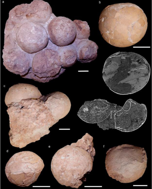
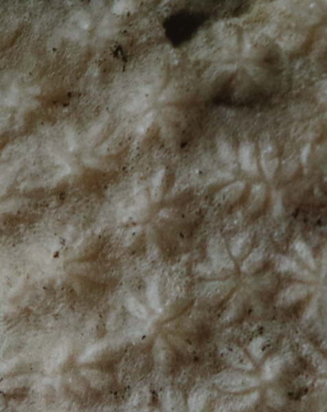
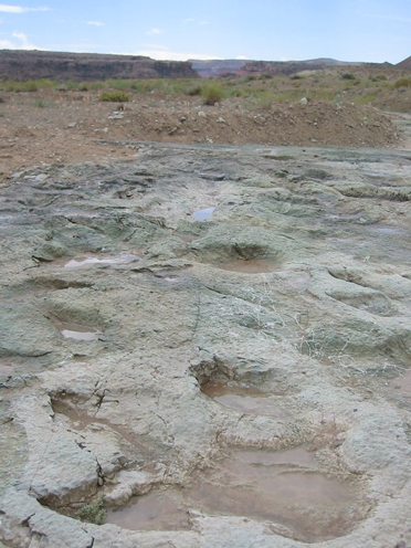
Images of selected titanosaurian eggs and egg-clutches collected from the Late Cretaceous Serra da Galga Formation (Bauru Group) at Ponto Alta nesting site, Uberaba Municipality, Minas Gerais State, Brazil. CPPLIP 1798, (a) the best-preserved recovered egg-clutch, bottom view. CPPLIP 1801, (b) isolated egg, with accompanying tomographic slice, showing thickness of the shell and its sedimentary fill. Specimen number CPPLIP 1799, (c) egg-clutch with accompanying tomographic slice, showing thickness of the shell, shells collapsed and its sedimentary fill. The specimen number CPPLIP 1800, (d and e) two eggs found associated. Specimen number CPPLIP 1804 (f) isolated partial egg. Note scale bars 5 cm. Picture credit: Fiorelli et al.

Picture credit: Fiorelli et al

Nesting in Colonies and Evidence of Breeding-site Fidelity

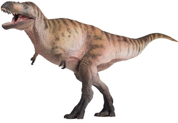
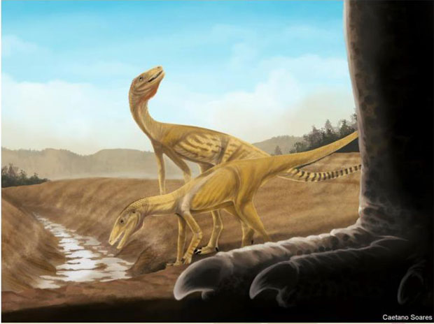
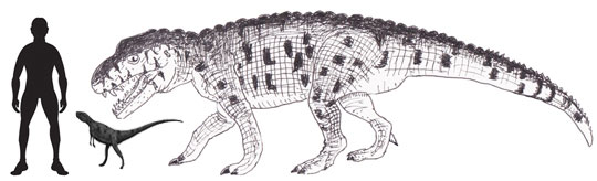
The Titanosauria were a highly successful clade of long-necked, long-tailed herbivores that were globally distributed during the Cretaceous and survived up until the very end of the Mesozoic. Some of the biggest terrestrial animals known to science are members of this clade, dinosaurs such as Patagotitan, Argentinosaurus and Notocolossus. All three of these titanosaurs lived millions of years before the Lafarge Quarry eggs were laid and they lived much further south (higher palaeolatitude).

Patagotitan scale drawing.

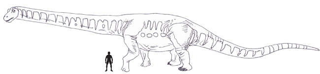
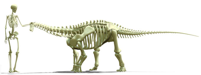
A scale drawing of the giant titanosaur Patagotitan mayorum commissioned by Everything Dinosaur. This huge dinosaur was estimated to have measured around 35 metres when its fossil bones were initially examined. When Patagotitan was formally named and scientifically described its body size and weight was reduced slightly. However, a reconstructed, life-size skeleton was built measuring 37.2 metres in length. Titanosaurs are amongst the largest terrestrial animals known to science. Picture credit: Everything Dinosaur.

Picture credit: Everything Dinosaur

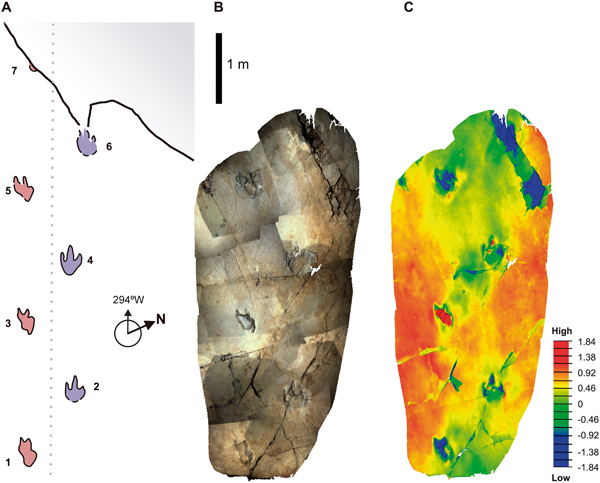
Numerous titanosaur nesting sites are known (Spain, Romania, France, India and most notably Argentina), writing in the academic journal “Scientific Reports”, the researchers report the first titanosaur nesting site from the Late Cretaceous of Brazil (Maastrichtian faunal stage). The fossil material consisting of several egg-clutches, partially preserved, isolated eggs and many eggshell fragments were found in two distinct levels, approximately two metres apart.

This discovery adds further support to the theory that these large herbivores nested in colonies and had preferred locations for their nesting sites, what is often referred to as breeding-site fidelity.

To read a related blog post about breeding-site fidelity (philopatry) identified in South American titanosaurs: Two New South American Titanosaurs Described.

Brazilian titanosaur nesting site

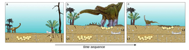
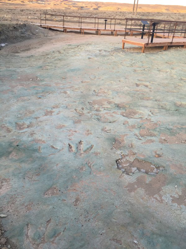
The titanosaur egg fossils were found in two distinct layers (L1 and L2) approximately two metres apart. This suggests that this area was a preferred nesting site for titanosaurs. This is the first confirmed dinosaur nesting area found in Brazil. The eggs attributed to titanosaurs also represent the most northerly titanosaurian nesting site known from South America. The discovery of nests located at different levels indicates that titanosaurs returned regularly to preferred nesting areas. Picture credit: Fiorelli et al.

Picture credit: Fiorelli et al

Which Titanosaur Species?

Isolated and fragmentary remains of titanosaur eggs had previously been reported from Brazil, but the Lafarge Quarry specimens provide unambiguous proof of titanosaur nesting in Brazil. As no embryos have been found in association with the fossilised eggs, it is not possible to comment on the specific genus involved.

However, the researchers state that between the Santonian to the Maastrichtian faunal stages of the Late Cretaceous (86 to 66 million years ago) only derived titanosaurs such as saltasaurids and colossosaurians are recorded from South America. It is likely that the fossils associated with the nesting site represent an unknown genus.

Patagotitan skeleton on display.

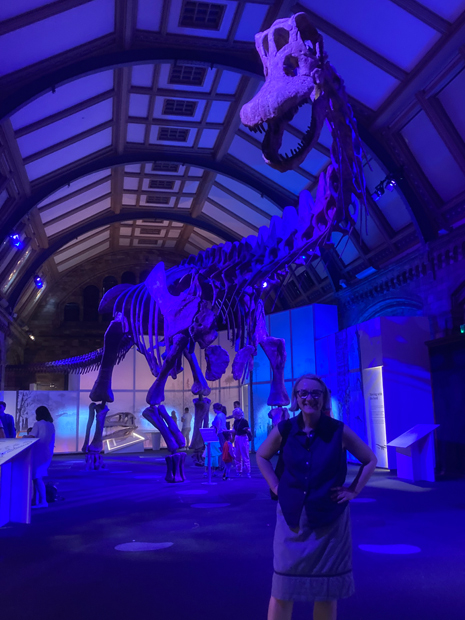
Sue from Everything Dinosaur poses in front of the colossal Patagotitan skeleton which is being exhibited at the Natural History Museum (London). Picture credit: Everything Dinosaur.

Picture credit: Everything Dinosaur

During the Late Cretaceous the Ponte Alta District would have been located at around 26 degrees south of the Equator, putting it on a similar palaeolatitude of titanosaur nesting sites found in India. Previously, South American titanosaur nesting sites have been recorded between 33 to 47 degrees south of the Equator. This latitudinal difference could also influence the distribution of species on the Gondwana continent. The palaeoclimatic variation and geological characteristics could result in a difference in nesting time between titanosaurs living at different latitudes.

How Did Titanosaurs Build Nests?

The Lafarge Quarry site indicates colonial nesting which also suggests that these herbivorous dinosaurs moved around in herds. It is likely that Ponte Alta District titanosaurs scooped out pits in the soft sand using their back legs and then laid their eggs in clutches, before burying them and perhaps covering the nest with vegetation to help incubation.

To read an article about the naming of Austroposeidon magnificus the largest dinosaur from Brazil described to date: Brazil’s Biggest Dinosaur.

The scientific paper: “First titanosaur dinosaur nesting site from the Late Cretaceous of Brazil” by Lucas E. Fiorelli, Agustín G. Martinelli, João Ismael da Silva, E. Martín Hechenleitner, Marcus Vinícius Theodoro Soares, Julian C. G. Silva Junior, José Carlos da Silva, Élbia Messias Roteli Borges, Luiz Carlos Borges Ribeiro, André Marconato, Giorgio Basilici and Thiago da Silva Marinho published in Scientific Reports.

The award-winning Everything Dinosaur website: Dinosaur Models.

22 03, 2022

Earliest Jaws in Evolutionary Trade-Off According to New Research

By |2024-10-30T10:02:57+00:00March 22nd, 2022|Categories: Adobe CS5, Dinosaur and Prehistoric Animal News Stories, Main Page, Palaeontological articles, Photos/Pictures of Fossils|0 Comments

Most vertebrates have jaws, specialised anatomical features for capturing and processing food. A newly published study suggests that the earliest jaws to evolve were caught in a trade-off between maximising their strength and their bite speed.

Dunkleosteus on display. A placoderm parent?

A spectacular Dunkleosteus exhibit. Dunkleosteus was one of 48 Placodermi genera whose jaws were examined in this study. Mathematical models of potential jaw shapes were compared against the actual fossil record. Jaw evolution has been constrained to shapes that have the highest possible speed and strength. Picture credit: Everything Dinosaur.

Picture credit: Everything Dinosaur

The Evolution of Jaws in Vertebrates

Jawed vertebrates dominate modern ecosystems emphasising the importance of jaw development in the evolutionary history of the vertebrates. Over 99% of all living vertebrates have jaws and the very first jaws evolved in prehistoric fish over 400 million years ago. These jaws evolved from gill arches, anatomical structures in fish that support their gills.

How jaws developed has been well documented, for example, Everything Dinosaur has blogged about their evolution: Ancient Shark Provides Insight into Jaw Development, however, researchers led by scientists from the University of Bristol have explored this important topic using a mathematical model to examine the early evolution and radiation of jaw types.

Megamastax amblyodus Illustration

Megamastax terrorises a group of jawless fish. The sarcopterygian Megamastax from the Late Silurian, a large, predatory fish with a powerful jaw attacking a group of jawless fish. The evolution of jaws probably led to the demise of the Agnatha (jawless fish) with only the Hagfish (Myxini) and the Lamprey (Hyperoartia) as the only living examples of this once extremely specious Infraphylum.  Picture credit: Dr Brian Choo.

Picture credit: Dr Brian Choo

The Development of a Novel Mathematical Model to Explore Jaw Development

Writing in the academic publication “Science Advances”, the researchers collected data on the shapes of fossil jaws and developed a mathematical model to assess their characteristics. The mathematical model produced permitted the scientists to extrapolate an extensive range of theoretical jaw shapes that could have been explored by the first evolving jaws. These theoretical jaws were tested for their strength – the amount of force they could endure without breaking, and their speed – how efficiently they could be opened and closed.

Jaw speed and jaw strength are two traits that conflict with each other. For example, really strong, powerful jaws like those associated with the giant placoderm Dunkleosteus would probably have been quite slow to open and close. Increasing bite force usually means a decrease in jaw speed.

By comparing the fossil record with the theoretical jaw shapes predicted by the mathematical model, the researchers deduced that jaw evolution has been constrained to shapes that have the highest possible speed in relation to their strength. Specifically, the jaws of early members of the Gnathostomata (jawed vertebrates) in the dataset were extremely optimal, and some groups evolved away from this optimum over time. These results suggest that the evolution of biting was very quick.

Studying the Earliest Jaws Using Mathematics

Bristol University PhD student William Deakin, the lead author of the paper explained:

“Jaws are an extremely important feature to gnathostomes – or jaw-mouths. They are not only extremely widespread, but almost all creatures that have them, use them in the same way – to grab food and process it. That’s more than can be said for an arm or a foot or a tail, which can be used for all sorts of things. This makes jaws extremely useful to anyone studying the evolution of function. Very different jaws from very different animals can be tested in similar ways. Here we have shown that studies on a large variety of jaws, using theoretical morphology and adaptive landscapes to capture their variety in function, can help shed some light on evolutionary questions.”

Scale drawing of a Coelacanth. What is a Coelacanth?

A scale drawing of a ceolacanth (Latimeria). he research team analysed the jaw shape of 121 extinct gnathostome taxa ranging in age from the Late Silurian to the end of the Devonian. The largest group of fossil fish studied were members of the Sarcopterygii (57 examples studied). The Sarcopterygii are the lobe-finned fishes an example of which is the extant Coelacanth (Latimeria). Picture credit: Everything Dinosaur.

Picture credit: Everything Dinosaur

Co-author of the study Professor Philip Donoghue (University of Bristol), added:

“The earliest jawed vertebrates have jaws in all shapes and sizes, long thought to reflect adaptation to different functions. Our study shows that most of this variation was equally optimal for strength and speed, making for fearsome predators.”

Emily Rayfield, who like Professor Donoghue is also a Professor of Palaeobiology at Bristol University and co-author of the paper, praised the new, insightful mathematical model developed by William Deakin commenting:

“The new software that Will developed to analyse the evolution of jawed vertebrates, is unique. It allows us to map the design space of key anatomical innovations, like jaws, and determine their functional properties. We plan to use it to uncover many more of the secrets of evolutionary history.”

Everything Dinosaur acknowledges the assistance of a media release from the University of Bristol in the compilation of this article.

The scientific paper: “Increasing morphological disparity and decreasing optimality for jaw speed and strength during the radiation of jawed vertebrates” by William J. Deakin, Philip S. L. Anderson, Wendy Den Boer, Thomas J. Smith, Jennifer J. Hill, Martin Rücklin, Philip C. J. Donoghue and Emily J. Rayfield published in Science Advances.

Visit the Everything Dinosaur website: Everything Dinosaur.

Go to Top