Pictures of fossils, fossil hunting trips, fossil sites and photographs relating to fossil hunting and fossil finds.
27
06, 2022
Frozen Baby Mammoth Discovered in the Klondike
Gold miners working at Eureka Creek in the Klondike Region of Yukon Province in Canada have discovered the frozen remains of a baby woolly mammoth. The calf, which is female is estimated to have lived around 30,000 years ago and it represents the best-preserved woolly mammoth specimen ever found in North America.

The baby mammoth identified as a female, is the best preserved Woolly Mammoth (Mammuthus primigenius) found to date in North America. It is thought to be around 30,000 years old. Picture credit: Yukon Government.
Picture credit: Yukon Government
“Big Baby Animal”
The discovery was made on June 21st, the Northern Hemisphere solstice and also appropriately, Canada’s National Indigenous Peoples Day. The Klondike gold fields lie within the Trʼondëk Hwëchʼin Traditional Territory. Trʼondëk Hwëchʼin elders have named the mammoth calf Nun cho ga, meaning “big baby animal” in the indigenous people’s (Hän) language.
Ice Age animal remains are quite commonly found in the Yukon area as they erode out of thawing permafrost, however, mummified remains complete with skin and hair are exceptionally rare.
Minister for Tourism and Culture, Ranj Pillai of the Yukon Territory Administration commented:
“The Yukon has always been an internationally renowned leader for ice age and Beringia research. We are thrilled about this significant discovery of a mummified woolly mammoth calf: Nun cho ga. Without strong partnerships between placer miners, Trʼondëk Hwëchʼin, and the Yukon government, discoveries like this could not happen.”
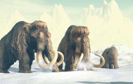
Woolly Mammoths (M. primigenius) an iconic animal of the Ice Age.
Vertebrate palaeontologist Dr Grant Zazula added:
“As an ice age palaeontologist, it has been one of my lifelong dreams to come face to face with a real woolly mammoth. That dream came true today. Nun cho ga is beautiful and one of the most incredible mummified ice age animals ever discovered in the world. I am excited to get to know her more.”
Comparisons with Lyuba
The discovery of the superbly preserved corpse will provide scientists with an opportunity to compare Nun cho ga with Lyuba, a mammoth calf discovered in Siberia back in 2007. Lyuba lived a few thousand years earlier than the Yukon mammoth (circa 41,800 years), researchers will have the opportunity to compare the genetic health of the mammoth population and plot any changes between the older Lyuba and Nun cho ga which lived, around 12,000 years later.
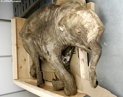
The 40,000-year-old baby mammoth Lyuba. Picture credit: Uppa/Photoshot (Daily Telegraph News).
Picture credit: Uppa/Photoshot (Daily Telegraph News)
The discovery of Nun cho ga is not the first woolly mammoth calf found in North America. In 1948, a partial mammoth calf, nicknamed Effie, was found at a gold mine in Alaska.
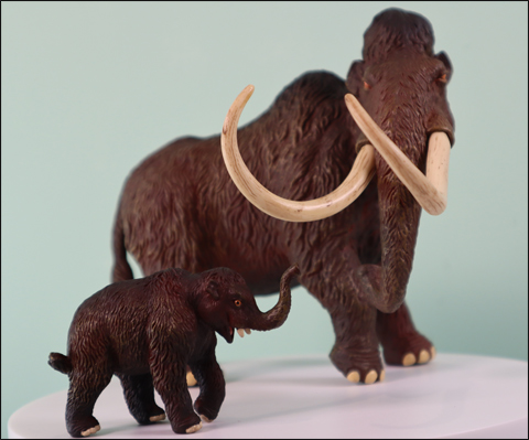
The CollectA Deluxe Woolly Mammoth model in 1:20 scale and the CollectA Prehistoric Life Woolly Mammoth calf. Picture credit: Everything Dinosaur.
Picture credit: Everything Dinosaur
The picture (above) shows a CollectA Deluxe Woolly Mammoth model and a CollectA Woolly Mammoth calf. This is a popular pair of prehistoric mammal models.
To view the range of CollectA scale models: CollectA Deluxe Prehistoric Life.
The CollectA Age of Dinosaurs Popular range: CollectA Age of Dinosaurs Popular Range.