Three-Dimensional Printers and Computerised Tomography Reveal Rare Fossils
3-D Printing Technology Used to Help Palaeontologists Reveal Fossils Still Inside Protective Plaster Jackets
More and more technology is being employed by palaeontologists these days and one of the latest tools in the armoury of a palaeontologist is a three-dimensional printer. In combination with computerised tomography (CT scans), a printer can produce an exact replica of a fossil, even one that is still buried deep inside its protective burlap and plaster jacket.
Computerised Tomography
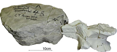
A team of German scientists have used a 3-D printer to create a copy of a fossil that may have been too delicate to remove from its jacket using conventional methods. This non-destructive technique opens up a whole new range of possibilities for palaeontology and other branches of the Earth sciences. The research team used CT scans to create a computerised image of an unidentified fossil bone which is part of the huge natural history collection of the Museum für Naturkunde, based in Berlin (Germany). The study has been published in the academic journal “Radiology”.
Field teams very often cover fossils that are being excavated in layers of sacking (burlap) and plaster. This is done to protect the fossil material in the field and in its subsequent transport back to the museum or the preparation lab. The plaster is carefully applied and then it is smoothed over by hand (surprising how many sharp edges can form on a plaster jacket), these lumps, some of which can weigh hundreds of kilogrammes, are then carefully removed and transported. Unfortunately, when it comes to removing the fossil from its jacket, this can be very tricky.
3-D Printers
Sometimes the plaster is so strongly bonded to the actual fossil bone that removing the plaster leads to damage of the fossil. Using the CT scans in combination with 3-D printers enables scientists to find out more information about a specimen without worrying about the removal of the surrounding sediment or plaster. The fossil is therefore very unlikely to be damaged using such technology.
The “scan and print” method as we call it at Everything Dinosaur is also very time efficient. It can take weeks, months even years to physically extract a fossil from its surrounding matrix.
The fossil specimen had been housed in a part of the Berlin museum that had been extensively damaged during World War II. Many of the specimens held by the museum were destroyed in bombing raids, others, like this specimen, ended up as unidentified objects, with researchers not sure as to what material the jackets may contain. Thanks to the application of this new technology, the research team were able to establish what was inside the plaster block and study the specimen that had been picked out by the powerful X-rays of the CT scan.
CT Scans
CT scans, otherwise known as CAT scans (computerised tomography) were originally conceived in the world of physics, but have applications in all sorts of other branches of science and elsewhere. This technology is used in medicine, engineering and of course in palaeontology. The X-rays from the CT scanner can distinguish fossil material from surrounding rock, sediment and plaster jackets as these materials all have different radiation absorption rates (different attenuation). This permits a computer to plot the data and produce an image in three-dimensions of any object such as a fossil inside a block of burlap wrapped stone.
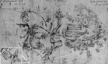
CT scans also provide palaeontologists with information regarding the placement and orientation of a fossil within a block, this can be extremely helpful when it comes to attempting a physical excavation. Information about the integrity of the fossil can be obtained, even possible fracture lines can be identified. The object in this study proved to be elements from the backbone, part of the Halberstadt excavations (Germany), a series of digs that took place between 1910 and 1927.
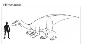
The vertebrae belongs to a Plateosaurus, a lizard-hipped, Late Triassic dinosaur that could have reached lengths in excess of ten metres. The bones measure approximately 21 centimetres in length and 17 centimetres in diameter at their widest part.
Picture credit: Radiology and RSNA
The application of this technology is helping the German scientists to sort out which of the unidentified parcels of fossil material belongs to which expedition. It turns out that this part of the museum that was damaged by bombing, housed fossil specimens collected in Germany and from Tanzania. The German led expeditions to Tanzania took place between 1909 and 1913, at the time, this was German East Africa.
The fossils collected by these two expeditions were mixed up. It now seems that 21st Century technology is going to help unravel a mystery surrounding fossilised bones of vertebrates that were collected in some cases more than one hundred years ago.
Picture credit: Everything Dinosaur
For models and replicas of Triassic dinosaurs and other prehistoric animals: CollectA Age of Dinosaurs Scale Models.
Once the CT images had been produced, the use of a computer allied to a 3-D printer was all that was required to reproduce a model of the object. Original drawings and diagrams from the expeditions were then used as a source of reference to help confirm the identity of the fossil material.
Picture credit: Radiology and RSNA
One of the authors of the research stated that the advances in three-dimensional printing technology means digital models of objects such as fossils can be transferred rapidly among researchers as endless copies can be made. Reproductions of fossils can be created from digital downloads, this will permit museums to share delicate, fragile fossils very easily. Very rare and important fossils such as those of early hominids could be made much more accessible, thus facilitating more research and study.


