A Very Poorly Duck-billed Dinosaur
A team of scientists, including a researcher based at the University of Manchester, have identified a case of septic arthritis in a dinosaur bone. This is the first documented case of this crippling condition to have been identified in the fossilised bones of a dinosaur. It is likely that the poor, unfortunate dinosaur, a hadrosaurid, struggled with painful joints for some time before it died.
The fossil bones, an ulna and radius (bones from the arm), were found in the Upper Cretaceous deposits of the Navesink Formation (New Jersey, USA). The strata were laid down in a near shore marine environment and dinosaur bones are exceptionally rare from these sediments, the ulna and radius were found fused together, but they were subsequently separated.
The ulna has a preserved length of 67.5 cm and the radius is slightly smaller at around 53.5 cm.
Bones from a Member of the Hadrosauridae?
The bones were compared to other hadrosaurid forelimb material and based on this analysis they were assigned to the Hadrosauridae (duck-billed dinosaurs), like many of the dinosaur fossil bones from eastern North America (Appalachia), these two bones could not be assigned down to the genus level. Writing in the open access journal “Royal Society Open Science”, the researchers refer to the specimens as coming from an indeterminate hadrosaurid.
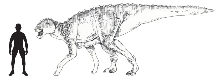
A Drawing of a Hadrosaurid Which May Have Been Similar to the Navesink Formation Specimen (NJSM GP11961)
Picture credit: Everything Dinosaur
Non-destructive Method of Viewing the Internal Structure of Fossil Bone
Historically, when palaeontologists have decided to assess the condition of a bone’s interior this has resulted in severe damage to the specimen. Prior to the use of X-ray microtomography (CT scans), the fossil bone would have had to be sectioned in order to reveal details of the inside. However, CT scans and other such non-invasive methods have enabled researchers to assess features inside the fossil in a non-destructive way. In this study, the ulna and radius (NJSM GP11961), were subjected to detailed cross-sectional CT scanning to assess the extent of the pathology present inside the fossil bones.
Pathology (Septic Arthritis) Identified in the Hadrosaurid Ulna
Picture credit: Royal Society Open Science
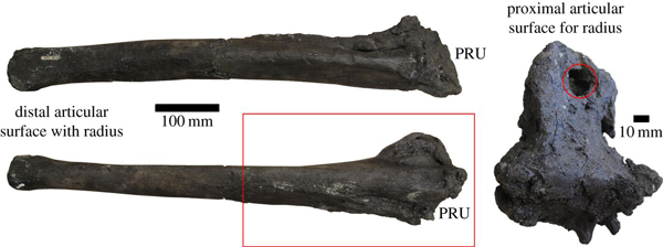
The picture above shows the hadrosaurid ulna (NJSM GP11961) in several views. The scientists identified a large area of roughened and remodelled bone towards the proximal articulation with the radius surface (PRU). Large lesions caused by the condition were found. The section of dinosaur bone in the red box shows that portion of the fossil that was subjected to X-ray microtomography.
X-ray microtomography Reveals the Extant of the Damage to the Ulna
Picture credit: Royal Society Open Science
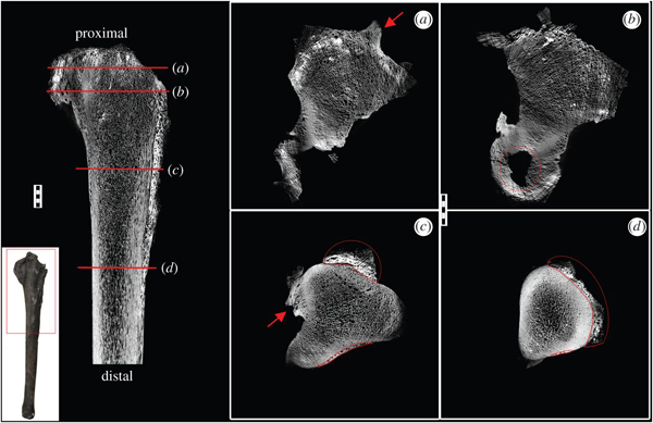
Note the scale bar is 1 cm. The lines labelled a to d in the image on the far left show the transverse sections on the ulna which correspond to the other four images shown. Abnormal, well-developed bony projections (enthesiophytes), are highlighted by the red arrows (images a and c). Necrosis (dead bone) is seen in b (circled in red) and remodelled bone growth is shown in both c and d (also outlined in red).
Comparing the Dinosaur’s Condition to Similar Conditions Found in Living Reptiles and Birds
The researchers, which included scientists from the University of Massachusetts and the New Jersey State Museum as well as Manchester University, looked at similar conditions seen in poultry and living reptiles and concluded that the abnormal bone growth, lesions and remodelled bone were likely caused by a form of osteoarthritis (arthritis in the bone).
Diagnosing Dinosaur Bone
Diagnosis was based on the erosion of the joint and highly reactive periosteal bone growth and fusion of the elements. This condition is caused by the loss of cartilage and it would have been very painful for the animal. Osteoarthritis in birds and reptiles is usually a consequence of disease, bacterial infection or trauma. The scientists were not able to state with any certainty how the condition came about, but at some point in this duck-billed dinosaur’s life it either had an accident, was attacked or caught an infection that led to this secondary condition. What the scientists were able to state was that this dinosaur lived with this painful condition for some time before it died.
Septic Arthritis Identified in the Hadrosaurid Radius
Picture credit: Royal Society Open Science
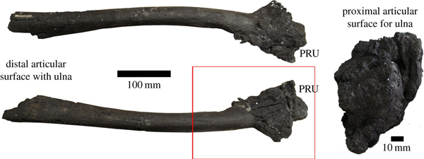
The picture above shows the hadrosaurid radius (NJSM GP11961), the red box indicates the area that was subjected to X-ray microtomography. The radius shows heavy reactive bone growth on the proximal articulation surfaces. In the picture, PRU (proximal articulation surface with the ulna), shows extensive pathological bone growth, described by the researchers as having a “cauliflower-like” appearance. The scientists noted that the distal articular surface with the ulna had eroded, most of the vertebrate fossils from the Navesink Formation tend to consist of isolated fragments, often quite heavily eroded due to taphonomy.
Septic arthritis is usually localised in reptiles, unlike in people, where the condition can spread throughout the body. However, as only the ulna and radius were found the extent to which the rest of the dinosaur’s body was affected is not known.
Visit the Everything Dinosaur website: Everything Dinosaur.




Leave A Comment