Studying the Growth Stages of Parareptiles
Juvenile Skull Specimen of Delorhynchus cifellii Helps in Growth Study
A team of scientists from the University of Toronto Mississauga (Canada), have analysed a total of seven different skulls of the parareptile Delorhynchus cifellii collected from the Early Permian fissure-fill deposits of Richard Spur, Oklahoma. This research, looking at the skulls which represent different growth stages of this primitive reptile, is helping palaeontologists to learn more about how the skulls of early tetrapods changed as they grew. It may also provide a valuable insight into how temporal fenestrae (holes in the skull), of other types of vertebrate evolved.
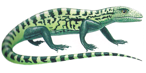
Delorhynchus cifellii
An Illustration of a Typical Early Reptile
Picture credit: Everything Dinosaur
For models and replicas of ancient Palaeozoic creatures: Prehistoric Animal Models.
Detailed Study of Cranial Ontogeny
Two species have been assigned to the genus Delorhynchu. The first D. priscus was named and described in 1962. In 2014, a second species was described D. cifellii, from a series of fossilised remains, including cranial (skull) material excavated from strata estimated to be around 275 million-years-old.
The researchers, which included Yara Haridy (Dept. of Biology, University of Toronto Mississauga), examined the partially articulated skull and jaw of a Delorhynchus cifellii believed to represent a juvenile (based on the size of the skull compared to other specimens and the lack of fully fused skull bones). This specimen probably represents an earlier growth stage (ontogenetic stage) of all the other known Delorhynchus skull material and as such, it helps provide a basis for a better understanding of how these ancient reptiles changed as they matured.
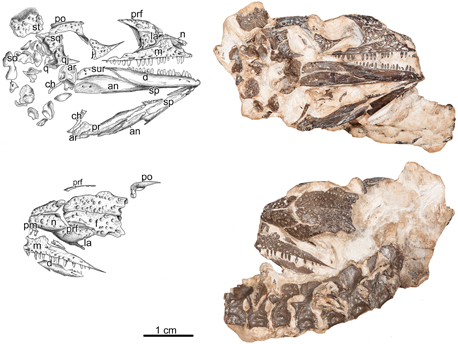
Views of the Delorhynchus cifellii Juvenile Skull Material Used in the Study
Picture credit: University of Toronto Mississauga
Analysing the Fossil Material
The photograph shows (above) a line drawing and image of the fossil material (right lateral view) and (below), a line drawing and image of the fossil material seen from above (dorsal view). The line of articulated vertebrae shown in the dorsal view do not belong to this individual. Scale bar = 1 centimetre.
Key
an, angular; ar, articular; ch, ceratohyal; d, dentary; f, frontal; j, jugal; la, lacrimal; m, maxilla; n, nasal; pal, palatine; pf, postfrontal; pm, premaxilla; po, postorbital; prf, prefrontal; q, quadrate; qj, quadratojugal; so, supraorbital; sp, splenial; sq, squamosal; st, supratemporal.
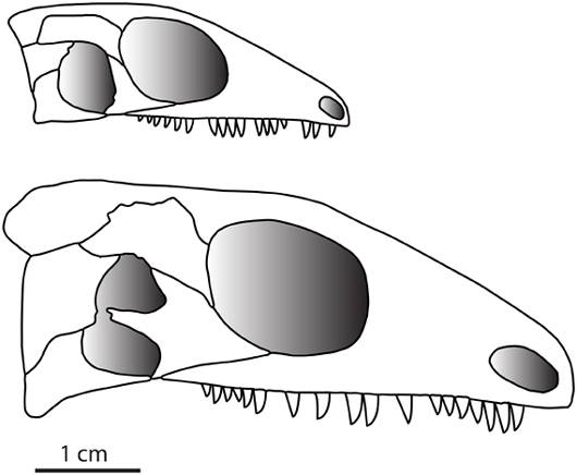
Comparisons between this juvenile and previously described specimens indicate that the size and shape of the temporal fenestra in Delorhynchus vary as the animal ages. These changes occur as the skull bones bordering the fenestra change shape. The growth series available for study, show that the jugal (cheek bone) becomes more robust as the reptile gets older and the proportionate size of the temporal fenestra is reduced. The scientists discovered that as Delorhynchus grew, the single, large fenestra seen in the skull of juveniles was gradually sub-divided into two smaller holes in more mature individuals.
Line Drawings Showing a Comparison between a Juvenile Delorhynchus Skull (Top) with a Mature Adult Delorhynchus (Bottom)

A comparison between the composite reconstructions of the youngest and most mature individuals in the Delorhynchus growth series.
Picture credit: University of Toronto Mississauga
The Fossil Record of Delorhynchus
The research team highlight the fact that the fossil record showing the growth of Delorhynchus is far from complete. They also point out that the largest specimen is not a fully mature individual. However, the data suggests that as the skull changed so the fenestra became much smaller and it would eventually be split into two small holes, due to the extension of the jugal bone.
The complete closure of the temporal fenestra may have occurred in very old animals, this cannot be ruled out, but the available skulls have provided a valuable insight into the possible growth trajectory of the fenestra of parareptiles. The scientists also speculate that the research on this anapsid (likely to have no fenestra in the skull when an adult), may help them to better understand the evolution of skulls in other anapsids, as well as diapsids (one pair of holes) and the synapsids (two pairs of holes).
The scientific paper: “Ontogenetic Change in the Temporal Region of the Early Permian Parareptile Delorhynchus cifellii and the Implications for Closure of the Temporal Fenestra in Amniotes” published in the on line academic journal PLoS One.
Visit Everything Dinosaur’s website: Everything Dinosaur.


