First Case of Septic Arthritis Diagnosed in a Dinosaur
Hadrosaur Had Septic Arthritis
Scientists writing in the Royal Society Open Science journal have identified the first diagnosed case of septic arthritis in a dinosaur. The unfortunate animal was a type of duck-billed dinosaur that roamed New Jersey (USA), some seventy million years ago. Identifying disease and injuries preserved in the fossil record (pathologies) permits scientists to gain valuable insights into the lives of long extinct creatures. The indeterminate duck-billed dinosaur may have suffered for many years with a debilitating injury.
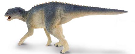
A Typical Hadrosaurid Hadrosaur
Picture credit: Everything Dinosaur
A Severely Damaged Elbow
The fossilised lower arm bones (ulna and radius) of a plant-eating, duck-billed dinosaur were excavated from sediments that form a component of the Upper Cretaceous exposures of the Navesink Formation (New Jersey). Both bones show malformation and deformed bone-growth, the result of some form of pathology. However, it was only when the team subjected the diseased part of the bones to an examination by a non-invasive technique, X-ray microtomography, could the research team, that included palaeopathologist Jennifer Anné, (University of Manchester), make a diagnosis.
The Diseased Ulna of the Indeterminate Hadrosaurid
Picture credit: Royal Society Open Science
A Damaged Dinosaur
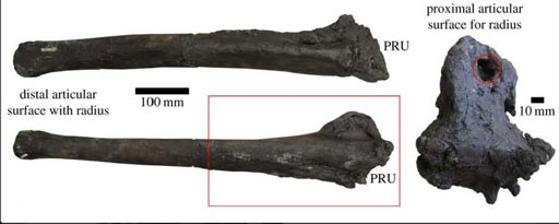
The photograph above shows the pathological ulna from an indeterminate species of duck-billed dinosaur. A substantial amount of bone re-growth can be seen on the end of the bone, the portion that is in articulation with the surface of the radius (PRU). The researchers describe the diseased bone as having a “cauliflower-like” appearance. The olecranon process that forms the elbow joint is very misshapen and damaged with large, prominent lesions (circled red). The red box in the diagram shows the portion of the ulna that was subjected to X-ray microtomography.
The research team conclude that this dinosaur would have been in pain and as a facultative biped it would have found movement difficult. However, bone re-growth suggests that this dinosaur lived for some time with this injury.
New Jersey and Dinosaurs
Although dinosaur fossils from the eastern coast of the United States are much rarer than those from the western USA, New Jersey is regarded by many scientists as the birthplace of American palaeontology.
The very first, scientifically described dinosaur discovery took place close to the town of Haddonfield in New Jersey. The bones of a large animal were excavated from a quarry and the eminent American scientist Dr Joseph Leidy (University of Pennsylvania), was given the task of studying them. He defined them as belonging to a member of the Order Dinosauria and erected the genus Hadrosaurus (H. foulkii).
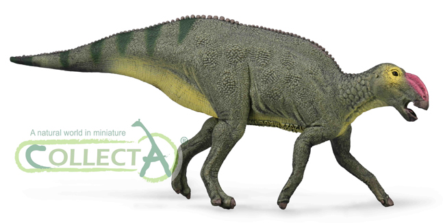
A Model of a Hadrosaurus (CollectA H. foulkii)
To view the CollectA range of prehistoric animal models: CollectA Prehistoric Life Models.
A statue of “Haddy” the Hadrosaur can be seen in Haddonfield, commemorating the discovery of “America’s first dinosaur.”
To read more about “Haddy” the Hadrosaur: A Hidden Gem (Dinosaur Statue).
Septic Arthritis
Having analysed the bones, the most likely explanation for the damaged bone is a form of osteoarthritis, a condition affecting movable joints by deterioration of articular cartilage, bone spur formation and this leads to considerable bone re-growth and remodelling. Such conditions are usually localised in extant reptiles, whereas in humans, these conditions can spread throughout the body.
It is worth noting that osteoarthritis in extant birds and reptiles is usually associated with other, contributory factors such as trauma, disease or infection. Based on the images generated by the X-ray scans, the researchers began to eliminate the possible causes such as osteomyelitis (the bone itself is infected) and finally concluded that the pathology probably represents septic arthritis (infected cartilage affecting the surrounding bone tissue).
What Caused the Injury?
If the septic arthritis was brought on by an injury then this begs the question, what caused the injury in the first place? Sadly, the seventy-million-year-old dinosaur arm bones don’t provide any clues.
Jennifer Anné commented:
“It could have started out that it did have arthritis. It could have gotten a cut, or broken that joint, and then had an infection. It’s a hard-knock life for any wild animal.”
X-ray Microtomography Scans (Longitudinal and Transverse Scans) Showing Disease Presence in the Ulna
Picture credit: Royal Society Open Science
Scanning the Hadrosaurid Ulna
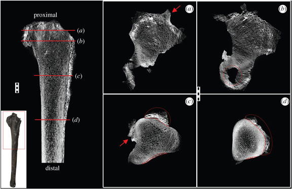
The picture above shows the scans of the hadrosaurid ulna in various views (a-d). The area scanned is shown on the picture on the white background in the lower left portion of the image. Locations for the transverse sections (a-d) are indicated by red lines on the longitudinal section. Reactive bone growth can be identified and is circled in pictures c and d. Abnormal bony projections at the attachment sites for ligaments (enthesiophytes) can be seen in a and c (indicated by red arrows). These abnormal bony growths are a sign of stress. Dead bone (necrosis) can be seen along the proximal articulation surface and is highlighted by a red circle in picture b. Scale bar for all images ten mm.
Dr Anné stated that the use of non destructive and non-invasive techniques such as X-ray microtomography is having a big effect on palaeopathology.
She added:
“As a result, how we’re approaching diagnosing is changing, it’s letting us look at more individuals, so we have a higher chance of finding things.”


