Beautiful Fish Fossil Illuminates Vertebrate Brain Evolution
A team of international scientists including researchers from the University of Birmingham have published a paper on the brain and cranial nerves of fish that lived approximately 319 million years ago. The team’s findings are shedding light on vertebrate brain evolution.
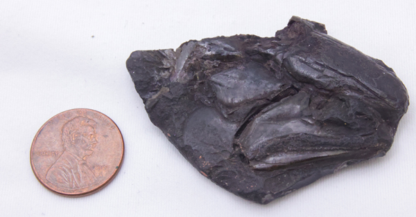
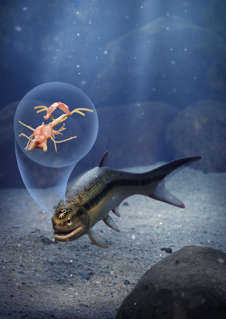
The Late Carboniferous (early Pennsylvanian subperiod), fish fossil was discovered in a layer of soapstone adjacent to a coal seam at the Mountain Fourfoot coal mine in Lancashire and the specimen was first scientifically described in 1925. The fish, named Coccocephalus wildi, would have measured around 20 cm in length and it lived in what was an ancient estuary. It is only known from this single fossil and only the skull and jaws were recovered.

Vertebrate Brain Evolution
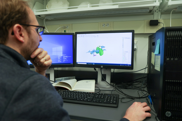
Coccocephalus was a member of the Class Actinopterygii, also known as the ray-finned fishes. The skull fossil was sent on loan from Manchester Museum to the University of Michigan and subsequent CT scans of the skull revealed the surprising discovery of the intact brain and associated nerves.
Senior author Sam Giles, (University of Birmingham), commented:
“This unexpected find of a three-dimensionally preserved vertebrate brain gives us a startling insight into the neural anatomy of ray-finned fish. It tells us a more complicated pattern of brain evolution than suggested by living species alone, allowing us to better define how and when present day bony fishes evolved.”

Rapidly Buried
When the fish died, it was probably buried rapidly in sediment containing very little oxygen. The lack of oxygen prevented the soft brain tissue from decaying. Whilst brain cases can reveal the shape and structure of vertebrate brains, this remarkable fossil preserved the brain tissue of a prehistoric fish.
Soft tissues such as the brain normally decay quickly and very rarely fossilise. But when this fish died, the soft tissues of its brain and cranial nerves were replaced during the fossilisation process with a dense mineral that preserved, in astonishing detail, their three-dimensional structure.
This discovery provides palaeontologists with a window into the evolution and development of the brains of ray-finned fishes, a highly successful group of back-boned animals estimated to represent more than fifty percent of all living vertebrate species.

A study of the jaws and teeth of C. wildi suggest that it was carnivorous, likely feeding on small invertebrates. The CT scans revealed that the brain had bilateral symmetry, like the brains of modern ray-finned fishes, but significantly, the brain of Coccocephalus folds inward, unlike in all living ray-finned fishes, in which the brain folds outward.
For figures and replicas of ancient prehistoric fish: Prehistoric Sharks, Dunkleosteus and Other Prehistoric Fish Models (PNSO).
The fossil captures a time before a signature feature of ray-finned fish brains evolved, providing an indication of when this trait evolved.
Co-author of the paper, published in the journal “Nature”, Matt Friedman (University of Michigan) explained:
“An important conclusion is that these kinds of soft parts can be preserved, and they may be preserved in fossils that we’ve had for a long time—this is a fossil that’s been known for over 100 years.”
Everything Dinosaur acknowledges the assistance of a media release from the University of Birmingham in the compilation of this article.
The scientific paper: “Exceptional fossil preservation and evolution of the ray-finned fish brain” by Rodrigo T. Figueroa, Danielle Goodvin, Matthew A. Kolmann, Michael I. Coates, Abigail M. Caron, Matt Friedman and Sam Giles published in Nature.

