
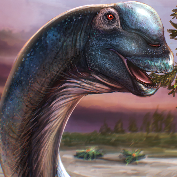
Future Marine Biologists Could Study Ichthyosaur Biology
Over the last two decades or so, a number of remarkable discoveries concerning soft tissue preservation within the fossil record have been made. Work pioneered by the remarkable Professor Mary Schweitzer (North Carolina State University), has led to evidence of dinosaur blood and traces of molecular biomarkers that represent preserved proteins such as collagen.
It may seem extraordinary, that such delicate evidence of organic remains can be preserved over huge amounts of time, but there is a growing body of data that indicates that such soft tissue preservation might be more common than previously thought. Under exceptional preservational circumstances soft tissue remnants could persist at the molecular level. A team of scientists led by researchers from Curtin University (Western Australia), have found red blood cells, collagen, white blood cells, platelet-like structures and cholesterol from the back-bone of an Early Jurassic ichthyosaur.
In the future, marine biologists could study the biology of a long extinct marine reptile…
Study Finds Soft Tissue Preservation in an Early Jurassic Marine Reptile
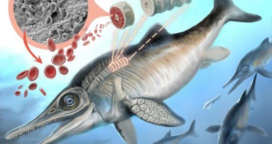
Traces of organic material preserved in an ichthyosaur.
Picture credit: Curtin University
Carbonate Concretion Gives Up Its Secrets
The researchers, led by organic geochemist John Curtin, analysed a dorsal vertebra from a 182.7 million-year-old ichthyosaur fossil from the world-famous Posidonienschiefer (Posidonia Oil Shales), of south-western Germany. Although vertebrate remains tend to be disarticulated, a lack of oxygen on the sea bed (anoxic conditions) at this location led to exceptional preservation conditions, there were few organisms present to decompose organic remains.
These conditions, in conjunction with rapid burial in the sediment has permitted remarkable fossil preservation to take place. Ichthyosaur carcasses have been preserved with soft tissue outlines intact, showing dorsal fins and tails, ink sacs and body outlines of belemnites such as Passaloteuthis sp. have also been preserved, allowing palaeontologists to vastly improve their understanding of the biology of these marine creatures. The high organic material content of the sediments permitted the formation of numerous carbonate concretions, these concretions isolate fossil material contained therein and can promote exceptional preservation of fragile tissues and biomolecules.
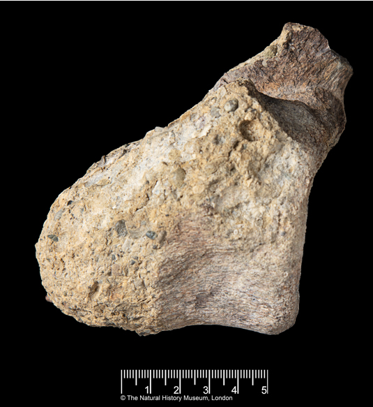
Ichthyosaur Fossil Jaw

A museum exhibit showing the jaw of a large ichthyosaur. Picture credit: Everything Dinosaur.
Picture credit: Everything Dinosaur
For models and replicas of ichthyosaurs and other marine reptiles: PNSO Age of Dinosaurs Models.
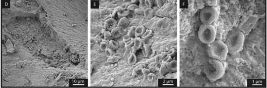
Stenopterygius Vertebra Studied
A single vertebra (back-bone) from a genus of ichthyosaur known as Stenopterygius was subjected to a range of microanalytical techniques, including scanning electron microscopy and microscopic calcite deposit removal at the molecular level using focused applications of acetic acid. Entombed inside the concretion, the internal structure of the vertebra, the spongy, trabecular bone, revealed biosignatures that represent the remnants of cholesterol, blood cells as well as straw-like structures that suggest the preservation of collagen fibres.
Scanning Electron Microscopy Reveals Collagen Fibres

Early Jurassic Stenopterygius vertebra reveals soft tissue preservation. (collagen fibres).
Picture credit: Curtin University/Scientific Reports
Co-author of the research paper, published this week in the academic journal “Scientific Reports”, Distinguished Professor Kliti Grice (Curtin University), explained:
“A carbonate concretion encapsulated the Early Jurassic period vertebra, forming a tight seal that helped protect its tissue and cellular remains from full decomposition.”
Compact and Trabecular Bone Studied
As well as the spongy, trabecular bone, the research team also examined the compact, cortical bone. Isotopic analysis of the cholesterol biomolecules is consistent with the view that ichthyosaurs dined on cephalopods and fish. Helping to reaffirm studies of coprolites and stomach cavity contents as to where in the marine food web these reptiles were situated.
Red Blood Cell Structures Identified in the Fossil Material
Red blood cell-like structures identified in fossil bone (ichthyosaur).
Picture credit: Curtin University/Scientific Reports
The Size of Red Blood Cells – Adaptations to Low Oxygen Levels
The doughnut shaped objects in the photograph (above), were identified as red blood cells. These cells were assessed to be up to five times smaller than those seen in extant animals. This finding led the researchers to propose the small size of these blood cell structures was related to the ichthyosaur’s evolutionary adaptation to environmental conditions – the lower oxygen levels associated with much of the Mesozoic.
Chloe Plet, a PhD student at Curtin University and a co-author of the scientific study stated:
“Ichthyosaurs evolved during a time when atmospheric oxygen levels were continuously low over a period of 70 million years. We propose that small red blood cells were favourably produced by the species to provide efficient oxygen transport and diffusion. For example, modern-day mammals living at elevated altitudes with lower oxygen levels make small and abundant red blood cells.”
Similarly sized red blood cells have been reported from dinosaurs (fossil material from the Upper Cretaceous), dinosaurs too, would have had to adapt to low atmospheric oxygen levels.
Understanding the Palaeobiology of an Ichthyosaur
The team conclude that the extraordinary preservation conditions associated with carbonate concretions could play a significant role in helping scientists to investigate the palaeobiology of long extinct species. Perhaps, one day in the future marine biologists will be able to study the biology of ichthyosaurs.
The scientific paper: “Palaeobiology of Red and White Blood Cell-like Structures, Collagen and Cholesterol in an Ichthyosaur Bone” by Chloé Plet, Kliti Grice, Anais Pagès, Michael Verrall, Marco J. L. Coolen, Wolfgang Ruebsam, William D. A. Rickard & Lorenz Schwark published by “Scientific Reports”.
Visit the Everything Dinosaur website: Everything Dinosaur.
link to the paper?