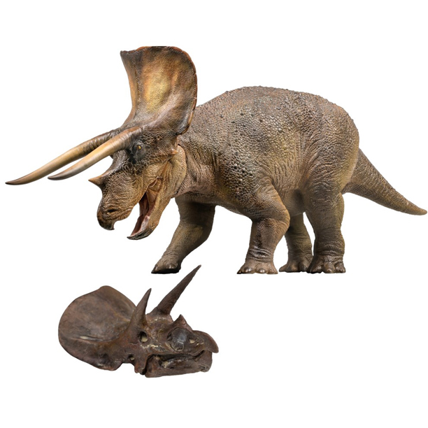
Visitors to the Berlin Naturkundemuseum (Germany) will be able to see an amazing Triceratops skull on display as part of an exhibition entitled “Dinosaurs! Age of the Giant Lizards”.
The impressive cranium, complete with horns and an imposing head shield measures two metres long and it was found in Lance Creek Formation deposits (Wyoming, USA) back in 2020. The fossil was discovered by an amateur fossil hunter and after preparation in Canada, the current owner Lars Fjeldsoe-Nielsen has lent the stunning specimen to the Museum für Naturkunde in Berlin.
The magnificent Triceratops skull on display in the “Dinosaurs! Age of the Giant Lizards” gallery at the Berlin Naturkundemuseum. Picture credit: Lukasz Papierak.
Horned Dinosaur Skull
The Triceratops specimen has been nick-named “Amalie” after the daughter of the owner. It is not known whether the skull fossil is from a female or male Triceratops. Both males and females sported neck frills and horns.
Numerous ornithischian dinosaurs are known from the Lance (Creek) Formation. The strata were deposited during the Maastrichtian faunal stage of the Late Cretaceous (69-66 million years ago). The fossils found in these rocks represent a diverse dinosaur dominated terrestrial fauna that thrived prior to the mass extinction event that saw the demise of the non-avian dinosaurs, including ceratopsians like Triceratops.
The new for 2022 PNSO Doyle the Triceratops 1:35 scale model comes complete with a scale model of a Triceratops skull.
The picture above shows a Triceratops model and skull, which is part of the PNSO Age of Dinosaurs series.
A spokesperson from Everything Dinosaur commented that they were unsure as to the Triceratops species that “Amalie” represented. They explained that both Triceratops horridus and an as yet, not fully described Triceratops species are associated with the Lance Formation.
Johannes Vogel, Director General of the Museum für Naturkunde Berlin thanked the owner for lending this wonderful specimen and stated:
“The Museum für Naturkunde Berlin would like to express its sincere thanks to Mr Fjeldsoe-Nielsen for this further generous loan. This will enable research museums like ours to get visitors excited about nature and explore the objects.”
The exhibition “Dinosaurs! Age of the Giant Lizards” is due to run until the end of the year.
Everything Dinosaur acknowledges the assistance of a media release from the Museum für Naturkunde Berlin in the compilation of this article.
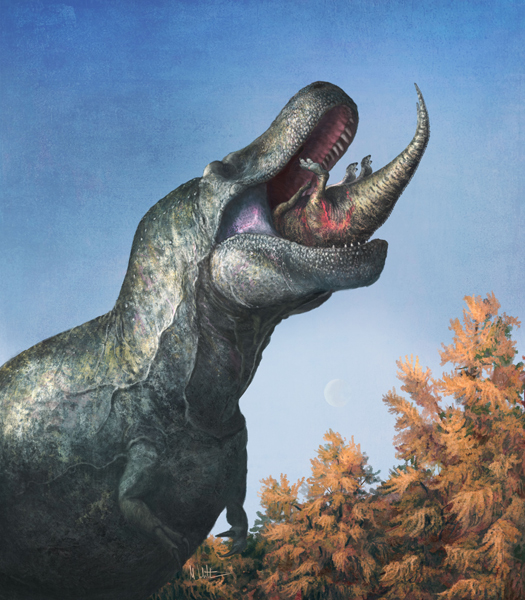
Large, predatory theropod dinosaurs are often portrayed as fierce-looking monsters, with huge and highly visible teeth. These teeth are visible over the jaw line when the meat-eating dinosaur’s mouth is closed. This is reminiscent of the appearance of modern crocodilians, which after all, are closely related to fellow archosaurs such as the theropod members of the Dinosauria. However, a new study suggests predatory dinosaurs had scaly, lizard-like lips. Even Tyrannosaurus rex had lips according to a new paper published in the academic journal Science.
A juvenile Edmontosaurus disappears into the enormous, lipped mouth of Tyrannosaurus. Picture credit: Mark Witton.
Tyrannosaurus rex Had Lips
The researchers including Dr Mark Witton (University of Portsmouth) and the study lead author Assistant Professor Thomas M. Cullen (Auburn University, Alabama) suggest that carnivores such as Tyrannosaurus rex did not have permanently exposed teeth. Films such as “Jurassic Park”, many palaeoartists and numerous model manufacturers have got it wrong. Instead, these dinosaurs had scaly lips, covering and sealing their mouths.
The debate as to whether theropod dinosaurs such as Giganotosaurus, Velociraptor, T. rex and Allosaurus had lips has gone on for some time. Did these dinosaurs have perpetually visible upper teeth that hung over their lower jaws and were therefore exposed and on view even with the jaw closed? The researchers suggest that dinosaurs such as Tyrannosaurus rex did not have a smile like a crocodile. Theropods possessed lips similar to those of lizards and the ancient reptile Tuatara, the only extant member of the Rhynchocephalia.
Detailed Study
In the most detailed study concerning the presence or otherwise of extraoral tissue in the Theropoda conducted to date, the researchers examined the tooth structure, wear patterns and jaw morphology of lipped and lipless reptile groups and found that theropod mouth anatomy and functionality resembles that of lizards more than the mouths of crocodilians.
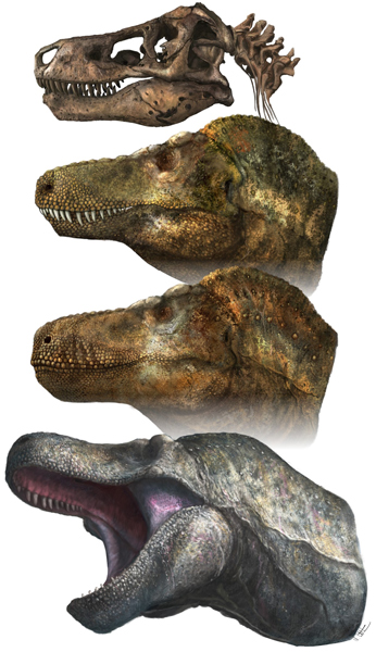
T. rex skull and head reconstructions. Picture credit: Mark Witton.
These lips were probably not muscular, like those of mammals. Most reptile lips cover their teeth but cannot be moved independently, a reptile can’t curl its lips back and snarl like a dog. They could not make the sort of movements that we might associate with our faces or that of other mammals.
Derek Larson, Collections Manager and Researcher in Palaeontology at the Royal BC Museum in Canada and a co-author of the study stated:
“Palaeontologists often like to compare extinct animals to their closest living relatives, but in the case of dinosaurs, their closest relatives have been evolutionarily distinct for hundreds of millions of years and today are incredibly specialised.”
The research team concluded that theropod teeth were extremely similar to the teeth of monitor lizards (varanids). The teeth are thought to have functioned in the same way, so perhaps monitor lizards such as the Komodo dragon (Varanus komodoensis) can be favourably compared to extinct animals such as theropod dinosaurs, even though the Varanidae as members of the Squamata, are only very distantly related to the Dinosauria.
Upending Popular Theropod Depictions
Co-author Dr Mark Witton (University of Portsmouth) explained:
“Dinosaur artists have gone back and forth on lips since we started restoring dinosaurs during the 19th century, but lipless dinosaurs became more prominent in the 1980s and 1990s. They were then deeply rooted in popular culture through films and documentaries — Jurassic Park and its sequels, Walking with Dinosaurs and so on. Curiously, there was never a dedicated study or discovery instigating this change and, to a large extent, it probably reflected preference for a new, ferocious-looking aesthetic rather than a shift in scientific thinking. We’re upending this popular depiction by covering their teeth with lizard-like lips. This means a lot of our favourite dinosaur depictions are incorrect, including the iconic Jurassic Park T. rex.”
The Implications of “Tyrannosaurus rex Had Lips”
The results of the study, found that tooth wear in lipless animals was markedly different from that seen in carnivorous dinosaurs and that dinosaur teeth were no larger, relative to skull size, than those of modern lizards, implying they were not too big to cover with lips.
Furthermore, the distribution of small holes around the jaws, which supply nerves and blood to the gums and tissues around the mouth, were more lizard-like in dinosaurs than crocodile-like. In addition, modelling mouth closure of lipless theropod jaws showed that the lower jaw either had to crush jaw-supporting bones or disarticulate the jaw joint to seal the mouth.
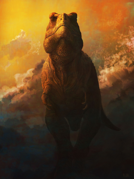
Tyrannosaurus rex bellowing with its mouth shut, like a vocalising alligator. Picture credit: Mark Witton
Kirstin Brink (Assistant Professor of Palaeontology at the University of Manitoba, Canada) and fellow co-author of the scientific paper commented:
“As any dentist will tell you, saliva is important for maintaining the health of your teeth. Teeth that are not covered by lips risk drying out and can be subject to more damage during feeding or fighting, as we see in crocodiles, but not in dinosaurs.”
Assistant Professor Brink added:
“Dinosaur teeth have very thin enamel and mammal teeth have thick enamel (with some exceptions). Crocodile enamel is a bit thicker than dinosaur enamel, but not as thick as mammalian enamel. There are some mammal groups that do have exposed enamel, but their enamel is modified to withstand exposure.”
Theropod Teeth are Not Oversized
Previously, it had been suggested that the teeth of predatory dinosaurs were just too big to be covered by lips. This study challenges that view and suggests that theropod teeth were not atypically large. Even the huge, banana-shaped teeth of tyrannosaurs are proportionally similar in size to living predatory lizards when the actual skull size is considered. Therefore, the researchers reject the hypothesis that theropod teeth were too large to be covered by lips.
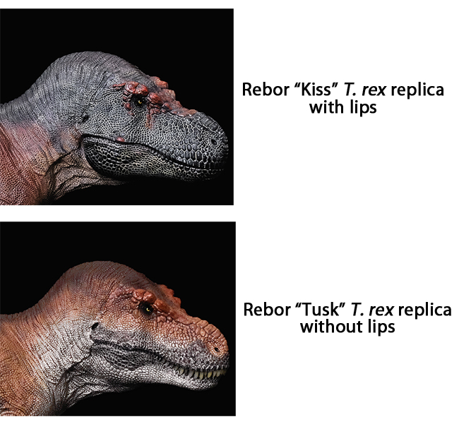
Model makers and figure manufacturers have created figures that reflect the current scientific debate about the presence or otherwise of lips in theropod dinosaurs. For example, Rebor recently introduced two new Tyrannosaurus rex figures “Kiss” being a lipped model, whereas the counterpart figure “Tusk” was lipless.
Some model manufacturers have reflected the current scientific debate by producing replicas with lips as well as lipless forms such as the recent Rebor “Kiss” and “Tusk” figures.
To view the range of Rebor figures and replicas in stock at Everything Dinosaur: Rebor Models and Figures.
Important Implications with Regards to Reconstructing Theropod Dinosaurs
This new study provides a new perspective on the “lips” versus “lipless” debate. It provides new insights into how scientists, artists and model makers reconstruct the soft tissues of dinosaurs and other prehistoric animals. This research provides information on how theropod dinosaurs fed, how they maintained their dental health as well as broader issues such as dinosaur ecology and evolution.
Dr Witton summarised the study stating:
“Some take the view that we’re clueless about the appearance of dinosaurs beyond basic features like the number of fingers and toes. But our study, and others like it, show that we have an increasingly good handle on many aspects of dinosaur appearance. Far from being clueless, we’re now at a point where we can say ‘oh, that doesn’t have lips? Or a certain type of scale or feather?’ Then that’s as realistic a depiction of that species as a tiger without stripes.”
The research team stress that their study does not say that no extinct animals had exposed teeth — some, like sabre-toothed carnivorous mammals, or marine reptiles and flying reptiles with extremely long, interlocking teeth, almost certainly did.
Everything Dinosaur acknowledges the assistance of a media release in the compilation of this article.
The scientific paper: “Theropod dinosaur facial reconstruction and the importance of soft tissues in paleobiology” by Thomas M. Cullen, Derek W. Larson, Mark P. Witton, Diane Scott, Tea Maho, Kirstin S. Brink, David C. Evans and Robert Reisz published in the journal Science.
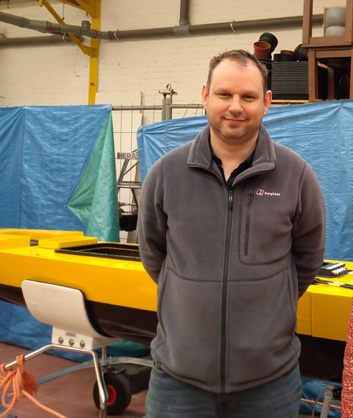
Plans are in place for an expedition to explore the seabed of the Adriatic for signs of early human settlement. Dr Simon Fitch, a geo-archaeologist at the University of Bradford is to embark on a mission to map submerged ice age landscapes and sunken settlements in what has been described as “a first of its kind”, scientific enquiry.
At the end of the month (March 2023), Dr Fitch will travel to Split in Croatia to begin a five-day survey of the Adriatic seabed using state-of-the-art underwater 3-D seismic sensors.
Dr Simon Fitch from the University of Bradford. Picture credit: University of Bradford/Simon Fitch.
Mapping Parts of the Adriatic and the North Sea
This expedition is the first of several that are being planned. Over the next five years, the researchers hope to map parts of the Adriatic and the North Sea. The North Sea being an area of particular interest to University of Bradford archaeologists as they have previously worked extensively on Doggerland, the huge tract of land that once linked Britain to continental Europe.
To read a recent article about research from the University of Bradford examining the impact of ancient Tsunamis on Doggerland settlements: Ancient Tsunamis Once Devastated Doggerland.
Between 24,000 and 10,000 years ago, global sea levels were around a hundred metres lower than they are today. This latest expedition is part of a long-term project to explore the archaeology of submerged human settlements.
The Life on the Edge Project
The Life on the Edge project is part of a UKRI future leaders fellowship for Dr Fitch, which last year attracted just over £1m in funding from UKRI, as well as £400,000 in-kind ship time from VLIZ (Flanders Marine Institute), and a PhD studentship from the University.
The University of Bradford’s Faculty of Life Sciences now has the largest submerged landscapes research group in the world and is one of the few places specialising in this exciting area of academic research.
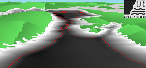
3-D image of the coastline of Croatia with the 14000-year-old coastline outlined in red. Picture credit: University of Bradford/Simon Fitch.
Commenting on the significance of the study, Dr Fitch stated:
“This is the first time anyone is going more than 500 metres from the coastline in the Adriatic to map the seabed. We know humans once lived on the land down there because trawlers regularly dredge up artefacts. This is about finding out who we are as a species and where we come from”.
An Incomplete Picture of Our History
Dr Fitch went onto explain that we have an incomplete picture of our own history. During the Late Palaeolithic (24,000 to 10,000 years ago), our planet was in the grip of an Ice Age and during this time we experienced the last “glacial maximum”, when sea levels were much lower than today, due to the amount of water stored in the ice caps and glaciers. More land around coasts would have been exposed and it is very likely that Stone Age people lived in these areas.
Dr Fitch added:
“We know most human populations like to live on the coastline, so it’s likely there were settlements on what is now the seabed. Our aim is to find evidence of those settlements and then recover the archaeology.”
Helping Renewable Energy Companies
Archaeologists from Bradford University along with collaborators from the University of Split and Flanders Marine institute (VLIZ), are working with commercial companies, who are already mapping the seafloor as they prepare to construct wind farms.
World map showing sea levels as they were during last glacial maximum, circa 22,000 years ago with yellow dots to show proposed wind farm activity. Picture credit: University of Bradford/Simon Fitch.
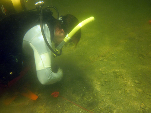
Powerful supercomputers installed at the University of Bradford are being used to process the huge volumes of data the expeditions will produce and The Life on the Edge Project has already attracted attention from other archaeologists based overseas. Dr Jessica Cook Hale (University of Georgia, USA), is to join the project.
The Search for Sunken Settlements
The academic is an experienced archaeologist with over two decades of research and field work behind her, including having dived underwater prehistoric sites in the Gulf of Mexico and the Atlantic coast.
Dr Jessica Cook Hale, (University of Georgia), who has joined the Life on the Edge project, diving a 5,000-year-old “midden heap” in the Econfina Channel site off the coast of Florida in August of 2015. Picture credit: Jessica Cook Hale.
Dr Cook Hale commented that she was excited to be joining this project and stated:
“Bradford is one of the few places doing this. I looked at this project from afar and wanted to be a part of it, so I’m thrilled to be joining the team. Carrying out geo-archaeology on submerged landscapes is really the only way to approach the problem of finding out about our prehistoric ancestors. As archaeologists, we’re naturally curious, we always want to ask, what came before?”
Training the Next Generation of Geo-archaeologists
One of the aims of the project team is to help recruit and train the next generation of geo-archaeologists.
The Life on the Edge project is an appropriate moniker, the team will be using cutting-edge mapping and computer technology and they will be exploring places that no archaeologists have explored before.
We wish the team every success with this intriguing venture.
Everything Dinosaur acknowledges the assistance of a media release from the University of Bradford in the compilation of this article.
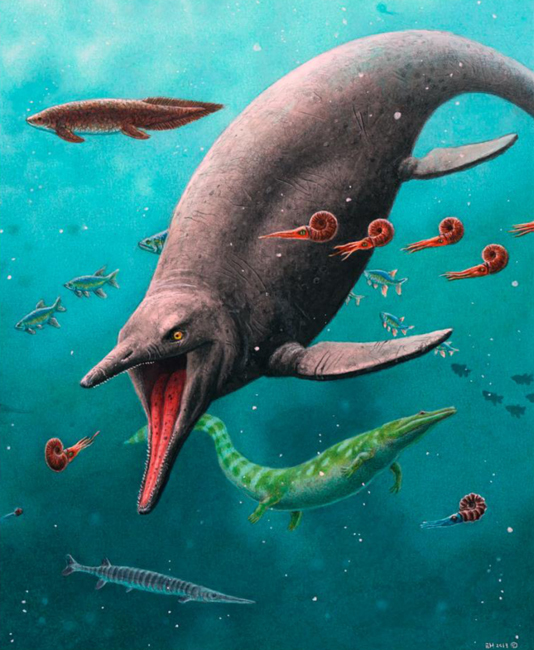
The earliest ichthyosaur fossil specimen discovered to date has been found on the Arctic island of Spitsbergen. The fossil represents a marine reptile that lived around 252 million years ago. The bones indicate that this animal was not a transitional form, but a fully adapted marine reptile.
Reconstruction of the earliest ichthyosaur and the 250-million-year-old ecosystem found on Spitsbergen. Picture credit: Esther van Hulsen.
Picture credit: Esther van Hulsen
Ichthyosaur Evolution
The evolutionary history of the ichthyosaurs remains contentious. No transitional forms representing land-dwelling tetrapods adapting to a marine habit have been found. However, small, basal ichthyosauriforms are known from the Lower Triassic of China, and the fossils of at least one, primitive Early Triassic, dolphin-shaped member of the Ichthyopterygia has already been described from Spitsbergen (Grippia longirostris).
Thanks to the work of a joint team of Swedish and Norwegian palaeontologists a fresh perspective on the origins of the “fish lizards” is provided by these newly described fossil bones.
An Ichthyosaurus model, typical of the dolphin-like, streamlined forms that existed during the Early Jurassic. Picture credit: Everything Dinosaur.
Ichthyosaurs were a highly successful, globally distributed group of marine reptiles. The evolved, a “dolphin-like” streamlined body and were active, nektonic predators surviving into the Late Cretaceous.
The first marine reptiles, such as the mesosaurs evolved during the Early Permian. The end-Permian mass extinction event devasted both terrestrial and marine faunas. The cataclysmic event was thought to have led to an evolutionary reset which permitted animals such as the Ichthyosauria to evolve, exploiting niches vacated after the extinction event.
Tetrapods (land-based vertebrates), invaded shallow coastal environments to take advantage of marine predator niches that were left vacant after the mass extinction event. Over millions of years, these early amphibious reptiles became more efficient at swimming and eventually modified their limbs into flippers, developed a ” dolphin-like” body plan, and started giving birth to live young (viviparity). With the evolution of viviparity, there was no need to come ashore in order to lay eggs, so the last ties these creatures had with a terrestrial existence was lost.
The newly described fossil material from Spitsbergen is helping to revise and re-write this previous hypothesis.
Fossil-bearing rocks on Spitsbergen that produced the earliest ichthyosaur remains. Picture credit: Benjamin Kear.
Picture credit: Benjamin Kear
Flower’s Valley Fossils
On western Spitsbergen a valley (Flower’s Valley), cuts deep into the surrounding mountains and provides access to Lower Triassic marine sediments, approximately 250 million years old. The rocks represent mud deposited at the bottom of an ancient sea and snow melt has gradually eroded the mudstone exposing rounded limestone boulders known as concretions. These objects are formed from limey sediments that coalesced around decomposing animal remains, subsequently preserving them in amazing, three-dimensional detail.
In 2014, the field team removed a number of concretions from the Flower’s Valley site. The rocks were taken back to the Natural History Museum at the University of Oslo for further study.
Scientists from The Museum of Evolution at Uppsala University have identified bony fish remains and bizarre “crocodile-like” amphibian bones, together with 11 articulated caudal vertebrae from an ichthyosaur.
Found in Rocks Thought to be Too Old for Ichthyosaur Fossils
Surprisingly, these tail bones occurred within rocks that were supposedly too old for ichthyosaurs. Also, the fossil bones do not represent a transitional form, but they show characteristics associated with geologically younger ichthyosaurs.
The vertebrae are identical to those of geologically much younger, larger-bodied ichthyosaurs, and even preserve internal bone microstructure showing adaptive hallmarks of fast growth, elevated metabolism and a fully oceanic lifestyle.
Computed tomography image and cross-section showing internal bone structure of vertebrae from the earliest ichthyosaur. Picture credit: Øyvind Hammer and Jørn Hurum.
Picture credit: Øyvind Hammer and Jørn Hurum
Dating the Surrounding Rock (Geochemical Testing)
Geochemical testing of the surrounding matrix dated the age of the fossils at approximately two million years after the end-Permian mass extinction. When the estimated timescale of marine reptile evolution is considered, this suggests the origins and early diversification of the Ichthyosauria took place during the Permian and prior to the Mesozoic Era.
These fossils suggest that the popular hypothesis of ichthyosaurs evolving to exploit niches vacated as a result of the end-Permian mass extinction is incorrect. Ichthyosaurs were present prior to the end of the Permian.
The discovery of the oldest ichthyosaur rewrites the popular vision of Age of Dinosaurs (Mesozoic Era), as the emergence timeframe of major reptile lineages. It now seems that at least some groups predated this landmark interval, with fossils of their most ancient ancestors still awaiting discovery in even older rocks on Spitsbergen and elsewhere in the world.
Everything Dinosaur acknowledges the assistance of a media release from the Uppsala University in the compilation of this article.
The scientific paper: “Earliest Triassic ichthyosaur fossils push back oceanic reptile origins” by Kear, B.P., Engelschiøn, V.S., Hammer, Ø., Roberts, A.J. and Hurum, J.H. published in Current Biology.
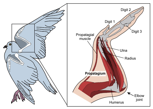
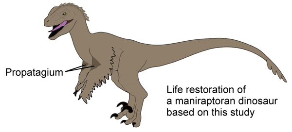
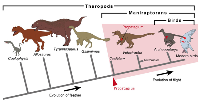
New research suggests maniraptoran theropod dinosaurs possessed a propatagium. The propatagium (pronounced pro-pah-ta-gee-um), is a soft tissue structure that joins the wrists and shoulders of volant birds. It helps with the wing flapping motion and provides a leading edge to the wing. Without this structure, birds could not fly.
If members of the Maniraptora, such as Therizinosaurus, Velociraptor, Oviraptor and troodontids had a propatagium on each arm, this would change how these dinosaurs are depicted. Many existing models and replicas would not be accurate and these figures would require updating.
Modern volant birds have a propatagium. A specialised wing structure, without which they would not be able to fly. The evolutionary origins of the propatagium remain uncertain, but new research led by scientists at the University of Tokyo (Japan), is helping to fill some of the gaps. By conducting a statistical analysis of the arm joints associated with the fossilised remains of some dinosaurs, the researchers have concluded that a propatagium was present in certain theropod dinosaurs on the dinosaur/bird evolutionary lineage.
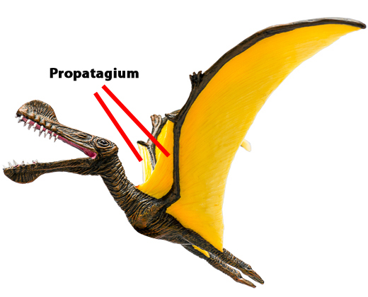
Propatagia are also known in other volant vertebrates – the bats and pterosaurs. These structures are examples of convergent evolution. Anatomical traits arising as animals adapt in similar ways to similar selective pressures.
A Tropeognathus pterosaur model with the propatagia highlighted.
Birds Evolved from Dinosaurs
Most scientists agree that birds evolved from maniraptoran dinosaurs. It therefore seems appropriate to look for avian traits within the Dinosauria, such as the presence of feathers, strong but light bones, and inner ears that help with balance and spatial awareness.
The University of Tokyo’s Department of Earth and Planetary Science wanted to try to see if evidence for the propatagium could be found in the non-avian dinosaur fossil record. The propatagium contains a muscle which connects the wrist to the shoulder and the research team set about trying to find evidence for this soft tissue structure in the fossilised remains of maniraptoran dinosaurs.
Co-author of the paper, published in the journal “Zoological Letters”, Associate Professor Tatsuya Hirasawa explained:
“It [the propatagium] is not found in other vertebrates, and it’s also found to have disappeared or lost its function in flightless birds, one of the reasons we know it’s essential for flight. So, in order to understand how flight evolved in birds, we must know how the propatagium evolved. This is what prompted us to explore some distant ancestors of modern birds, theropod dinosaurs.”
Theropod dinosaurs such as Giganotosaurus, Tyrannosaurus rex and Velociraptor had arms, not wings, although some theropods such as the dromaeosaurid Microraptor were capable of flight. If the researchers could find evidence of early examples of the propatagium within non-avian dinosaurs, they would gain a better understanding of how some Dinosauria gradually transitioned from having arms to evolving wings.
Unfortunately, a soft tissue structure such as a propatagium would only be preserved in exceptional circumstances. Hard, mineralised parts of the body such as bones have a far greater fossilisation potential. Perhaps the bones of fossilised dinosaurs could provide a clue?
Co-author of the study, Yurika Uno (University of Tokyo) explained:
“The solution we came up with to assess the presence of a propatagium was to collect data about the angles of joints along the arm, or wing, of a dinosaur or bird.”
Studying Joint Angles
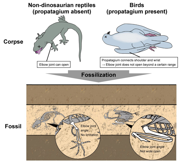
The presence or lack of a propatagium could be inferred by examining the angles of the joints in the arm in articulated fossil specimens. The way arm joints are articulated in fossils gives away the presence or absence of the propatagium structure. Thus the researchers could provide indirect evidence demonstrating the evolution of the avian wing structure.
The graduate student added:
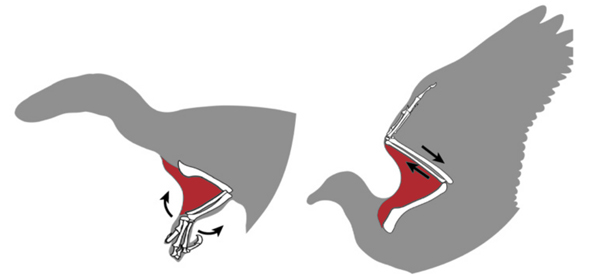
“In modern birds, the wings cannot fully extend due to the propatagium, constraining the range of angles possible between connecting sections. If we could find a similarly specific set of angles between joints in dinosaur specimens, we can be fairly sure they too possessed a propatagium. And through quantitative analyses of the fossilised postures of birds and nondinosaurs, we found the tell-tale ranges of joint angles we hoped to.”
The researchers postulate that the propatagium likely evolved in a group of dinosaurs known as the maniraptoran theropods. The Maniraptora clade is composed of coelurosaurian dinosaurs and is defined as including all birds and the non-avian dinosaurs that were more closely related to birds than they were to Ornithomimus velox.
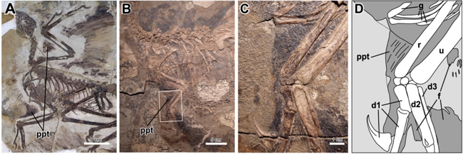
Close examination of the fossilised remains of the oviraptorosaurian Caudipteryx and the winged dromaeosaurian Microraptor indicate the presence of propatagia. The researchers suggest that they have found evidence for the presence of a propatagium in dinosaurs that existed prior to the evolution of flight in the maniraptoran lineage.
If maniraptoran dinosaurs had propatagia prior to the evolution of powered flight, then this raises an intriguing question. Why did the propatagium evolve? Why did these particular theropods evolve such a structure?
The University of Tokyo researchers are optimistic that by studying more fossils as well as embryonic development within extant vertebrates they might be able to provide some answers.
The team thinks some theropods might have evolved the propatagium not because of any pressure to learn to fly, as their forelimbs were made for grasping objects and not for flying. The propatagium originally had another purpose. It could be speculated that this “leading edge” of the arm evolved to help amplify visual intraspecific communication. Perhaps it evolved as a soft tissue structure used in display to demonstrate fitness for breeding and to win mates.
An enlarged surface area of the forelimb might have played a role in helping to shade eggs or perhaps play some other role in the brooding process.
Finding fossil evidence to support these suggestions is likely to prove difficult. However, if further studies demonstrate the presence of propatagia in the Maniraptora, it will change the way these types of dinosaurs are depicted.
Everything Dinosaur acknowledges the assistance of a media release from the University of Tokyo in the compilation of this article.
The scientific paper “Origin of the propatagium in non-avian dinosaurs” by Yurika Uno and Tatsuya Hirasawa published in Zoological Letters
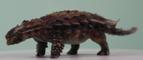
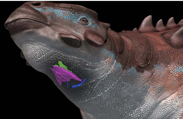
A team of scientists have been studying a Pinacosaurus larynx and have concluded that this armoured dinosaur was probably capable of producing a variety of sounds and calls.
A juvenile specimen of Pinacosaurus (P. grangeri), specimen number IGM100/3186, preserves a hyoid and two laryngeal elements (cricoids and arytenoids) in almost life articulation. From these remains the researchers have concluded that just like crocodilians and birds, Pinacosaurus was capable of producing a range of vocalisations. The calls may have had several functions, to alert others of a predator approaching, to threaten a predator, to define territory or to search for a mate. The sounds made by this ornithischian dinosaur may have been related to courtship, or perhaps helped to call offspring to their side.
Skull in ventral view (a) photograph by Michael D’ Emic and edited by Junki Yoshida. A 3-D reconstruction of the skull, jaws and hyolaryngeal apparatus in left oblique view (b). Crico-aryteniod joint of right cricoid in medial view (c). The joint of left arytenoid in dorsolateral view (d). Arytenoid position in glottal opening (e) and glottal closing in anterior views (f). Arytenoid position in glottal opening (g) and glottal closing in dorsal views (h). Abbreviations: afa, articular facet for arytenoid; afc, articular facet for cricoid; ap, arytenoid process; atr, atlas rib; caj, crico-arytenoid joint; lcb, left ceratobranchial; lcr, left cricoid; md, mandible; pm, premaxilla; pd, predentary; rar, right arytenoid; rcb, right ceratobranchial; rcr, right cricoid. Scale bars equal 1 cm. Picture credit Yoshida et al.
Pinacosaurus grangeri
Pinacosaurus (P. grangeri) is regarded as a basal member of the Ankylosaurinae subfamily of ankylosaurs. It is known from copious fossil material, and it is one of the most extensively studied of all the Late Cretaceous Thyreophora. Fossils are known from the Mongolia and China (Djadokhta Formation and the geologically older Alagteeg Formation).
A Pinacosaurus dinosaur model (PNSO). A study into their vocalisation has been published. Picture credit: Everything Dinosaur.
The image (above) shows a not-to-scale replica of Pinacosaurus (PNSO).
In tetrapods the voice box (larynx) has several functions. It plays a role in respiration, protects the airway to prevent food items becoming lodged and it has a function in vocalisation. Fossil preservation of the larynx in archosaurs is extremely rare. The Pinacosaurus fossil material (IGM100/3186) represents the oldest voice box known to science. It provides scientists with an opportunity to better understand the evolution of the larynx in non-avian dinosaurs.
The Pinacosaurus hyolaryngeal apparatus (tongue and voice box) in situ. A life reconstruction. Cricoid (purple), arytenoid (green), and ceratobranchial (blue) are depicted. Artwork by Tatsuya Shinmura.
Vocal Armoured Dinosaurs
Ossification of the cricoid and arytenoid is confirmed in Pinacosaurus, and it has been reported in Saichania, another Asian ankylosaurine. This configuration is also found in extant birds. The complex arrangement of the hyolaryngeal apparatus led the researchers to conclude that it did not simply function as a barrier to preventing food entering the trachea (airway protection). It was specialised for opening the glottis and possibly acting as a sound modifier.
The voice box of modern birds and crocodilians differs. In crocodiles and their close relatives it is the larynx that produces sounds. In birds, the larynx forms part of the vocal tract but they have a specialised organ (syrinx) located at the base of the trachea (wind pipe), that produces sounds.
Pinacosaurus – Shared Anatomical Characteristics
The researchers suggest that Pinacosaurus retained the same hyolaryngeal elements as found in crocodilians. However, Pinacosaurus shows many shared characters with birds in the arrangement and morphology of the larynx.
The authors of the scientific paper, which was published this month in “Communications Biology” (Junki Yoshida, Yoshitsugu Kobayashi and Mark Norell), propose that Pinacosaurus did not use the larynx as a sound source like non-avian reptiles. The larynx probably worked as a sound modifier as found in birds
Furthermore, the authors postulate that bird-like vocalisation likely appeared in non-avian dinosaurs before the evolution of the Aves (birds).
Article sourced from the open-access paper in Communications Biology.
The scientific paper: “An ankylosaur larynx provides insights for bird-like vocalization in non-avian dinosaurs” by Junki Yoshida, Yoshitsugu Kobayashi, Mark A. Norell published in Communications Biology.
It is widely accepted by palaeontologists that birds are descended from theropod dinosaurs. Their evolutionary lineage, the transition over time from the fast-running, agile, terrestrial Maniraptora to the birds we see today remains not fully understood. A new research project is being set up giving scientists the opportunity of tracing dinosaur footsteps to help them to better understand the evolutionary path of the avian dinosaurs.
A three-toed theropod footprint: Picture credit: Dr Peter Falkingham.
A £2.2 million GBP ($2.65 million USD) Research Project
A £2.2 million GBP ($2.65 million USD) research project funded by the European Research Council is being set up to permit scientists to study the evolution of the Dinosauria through their fossil tracks. The research project is to be led by Dr Peter Falkingham, a reader in vertebrate biology in the School of Biological and Environmental Sciences at Liverpool John Moores University.
Fossils of feathered dinosaurs and Mesozoic birds are known and have been extensively studied. Perhaps, one of the most intensively studied species in the entire fossil record is Archaeopteryx lithographica, a feathered theropod from the Upper Jurassic of southern Germany.
This five-year research programme, with its focus on studying theropod trace fossils, will provide a fresh perspective on the locomotion of the theropod/avian lineage.
Archaeopteryx fossil cast. Archaeopteryx is arguably one of the most extensively studied genera in the fossil record. This new research programme will focus on the locomotion of theropod dinosaurs. Picture credit: Everything Dinosaur.
Commenting on the scope of the study, Dr Falkingham explained:
“Fossil footprints are a direct record of motion in a way that skeletons can never be. I will use fossil footprints to explore the locomotor changes that took place as theropod dinosaurs evolved into birds.”
Creating a New Team
The plan is to establish a new team of post-doctoral scientists and technicians that will undertake advanced 3-D imaging of fossilised tracks and fossil skeletons. By combining trace fossils and body fossils in this way, the team hope to utilise kinematic and kinetic analyses to build an unprecedented view of footprint formation.
A simulated footprint (Guineafowl) mapped. Picture credit: Dr Peter Falkingham.
Tracing Dinosaur Footsteps
Limb motions of dinosaurs will be reconstructed using fossil tracks. Supercomputer simulations modelling every grain of a sediment responding to the indenting foot will be used to evaluate the reconstructed motions.
Dr Falkingham commented:
“These simulations will compute the forces occurring between foot and ground. These forces and motions will drive musculoskeletal biomechanical simulations that will shed light, not only on what the feet of dinosaurs were doing, but on how the whole limbs and even bodies of these enigmatic animals once moved. By sampling fossil tracks from around the world, spanning the 230 million years since theropods first appeared, this project will recover fossilised motions along the dinosaur-bird lineage.”
Mapping the locomotion of the avian lineage. Picture credit: Dr Peter Falkingham.
Extending our Knowledge About the Dinosaurs
Dr Falkingham added:
“The results should give us a unique view of locomotor evolution that cannot be recovered from bones alone.”
Everything Dinosaur acknowledges the assistance of a media release from Liverpool John Moores University in the compilation of this article.
For further information and to follow the progress of this research project, visit the website of Dr Peter Falkingham: Dr Peter Falkingham.
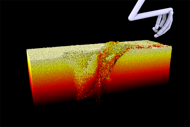
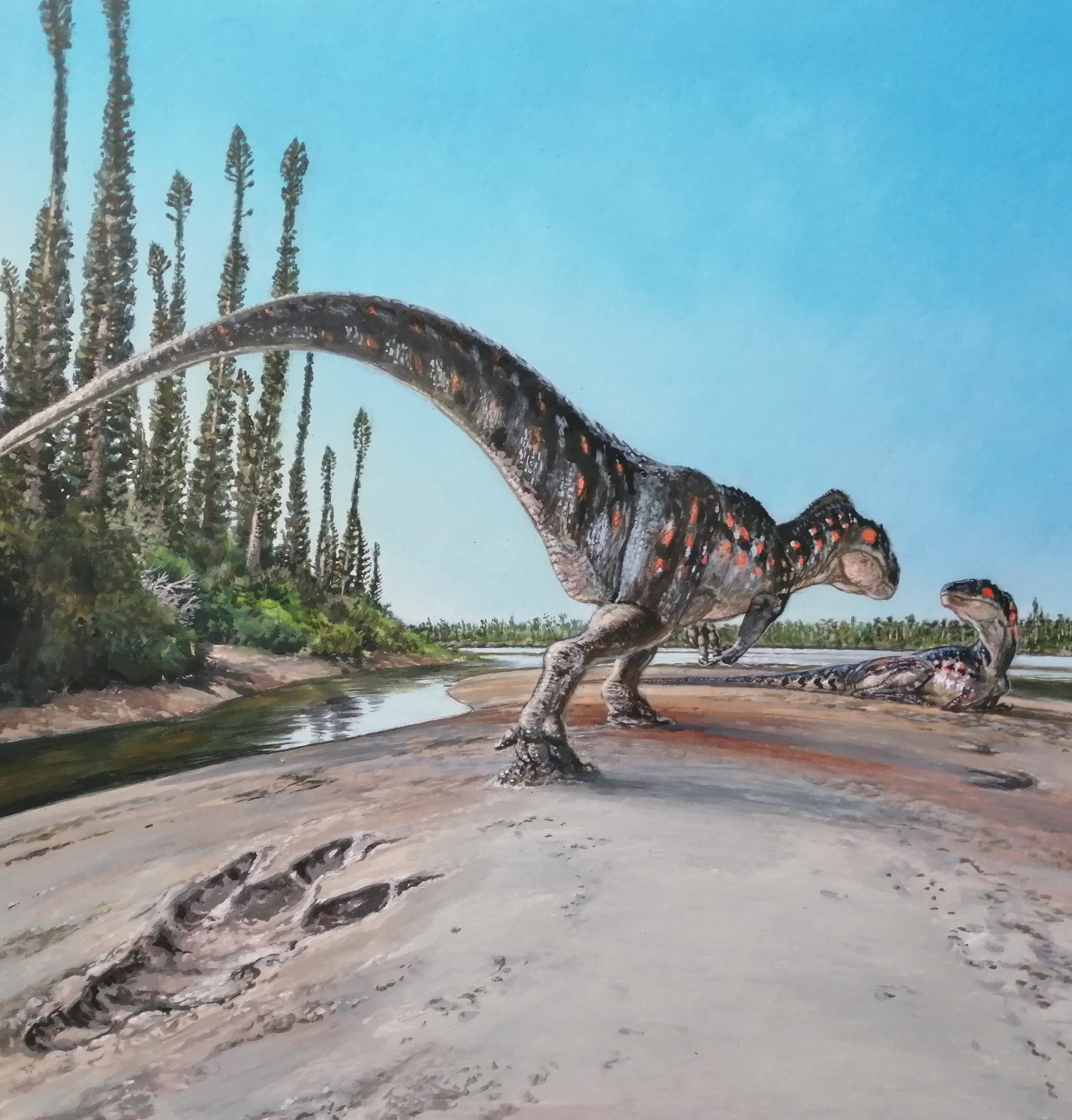
A giant, carnivorous dinosaur left an unusual footprint in soft sediment approximately 166 million years ago. Remarkably, the print has been preserved as a fossil, providing palaeontologists with yet more evidence to demonstrate the diverse, dinosaur dominated ecosystem which has been preserved in the rocks that comprise the Yorkshire coast.
Giant Dinosaur Footprint
The trace fossil measures around eighty centimetres in length, and it was probably made by a large theropod dinosaur (Megalosauridae).
The print was probably made by a large, carnivorous dinosaur similar to a Megalosaurus (Theropoda – Megalosauridae). Picture credit: James McKay.
Picture credit: James McKay
The Yorkshire Coast
The Yorkshire coast is renowned for producing some visually and scientifically significant fossils, including thousands of dinosaur footprints and tracks. A popular destination for professional palaeontologists and fossil fans, people come from far and wide to see what they can discover.
The three-toed (tridactyl) print is exceptionally rare and unusual. It appears to record the moment when a meat-eating dinosaur crouched down or rested.
Discovered by a Local Archaeologist
The print was discovered in April 2021 by Marie Woods, a local archaeologist. She was walking along the coast and found this amazing trace fossil by chance. Marie contacted local fossil experts to see if the print had already been recorded, but none of them were aware of the track she described.
Dr Dean Lomax, a vertebrate palaeontologist affiliated with The University of Manchester was contacted and asked to examine the fossil find.
Dr Lomax, a co-author of the study published in the Proceedings of the Yorkshire Geological Society commented:
“I couldn’t believe what I was looking at, I had to do a double take. I have seen a few smaller prints when out with friends, but nothing like this. I can no longer say that ‘archaeologists don’t do dinosaurs’. At the time of the discovery, it generated a lot of public interest and I was overwhelmed with the messages on social media from people around the globe.”
The large tridactyl print, the toes are on the right side of the photograph. The footprint was produced by a large theropod dinosaur and it measures approximately 80 cm in length. Picture credit: Marie Woods.
Picture credit: Marie Woods
An Extremely Significant Fossil Discovery
The footprint is one of only six similar prints to have been recorded in the area, the first of which was identified in 1934. This print is an extremely significant fossil discovery, not only are tridactyl prints rare, but this trace fossil is the largest found in Yorkshire to date.
Local geologist and lead researcher on the paper John Hudson explained:
“This important discovery adds further evidence that meat-eating giants once roamed this area during the Jurassic. The type of footprint, combined with its age, suggests that it was made by a ferocious Megalosaurus-like dinosaur, with a possible hip height between 2.5 and 3 metres.”
Dinosaurs of the British Isles
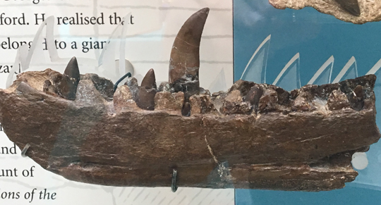
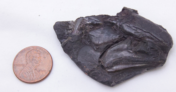
Megalosaurus (M. bucklandii), was the first dinosaur to be formally described (1824). Around a hundred different dinosaur genera have been described from fossils found in the British Isles. Such is the significance of dinosaur fossils from the UK, that Dr Lomax was inspired to write a book documenting the extensive dinosaur fossil discoveries that have been made on these islands.
The partial dentary for teeth associated with Megalosaurus bucklandii. Picture credit: Everything Dinosaur.
Picture credit: Everything Dinosaur
A Fragile Fossil
Photographs shared between the research team led them to conclude that the specimen was exceptionally fragile and likely to suffer further damage if it remained on the shoreline. Action was taken to rescue the fossil. The dinosaur trace fossil was expertly recovered by experienced fossil collectors Mark, Aaron and Shae Smith of Redcar.
As the rescue mission progressed it came to light that the print had been spotted five months previously, by Bob Taylor a local fossil collector who subsequently helped to write the research paper on the specimen.
Dr Lomax thanked Mark, Aaron and Shae for ensuring the safe recovery of the fossil and he stated:
“We’re incredibly grateful to Mark, Aaron and Shae for rescuing this important specimen and ensuring that it was saved for science. Now that the specimen has been studied, plans are in motion for it to go on public display, to spark the imagination of the next generation of fossil hunters.”
Donated to Scarborough Museum and Galleries
The fossil has been donated to Scarborough Museum and Galleries. Plans are in place to include this remarkable dinosaur in an exhibit, once conservation has been completed.
Dr Mike Romano (University of Sheffield), an expert on dinosaur tracks and other trace fossils, also co-authored the scientific paper. Dr Romano has spent more than two decades researching the dinosaur tracksites associated with the coast of Yorkshire.
He added:
“The east coast of Yorkshire is known as the Dinosaur Coast for very good reasons”
A huge number of dinosaur tracks, ranging in the thousands, have been discovered. As a result, this stretch of coastline is considered one of the best places in the world for dinosaur footprints. Although the first prints were documented in 1907, it was not until the 1980s that finds were being reported on a regular basis (by amateurs as well as professional geologists).
Twenty-Five Different Types of Track Described
Around twenty-five different types of footprints have been identified from the Jurassic strata exposed on the coast of Yorkshire. These prints and tracks demonstrate that during the Middle Jurassic a diverse, dinosaur-dominated ecosystem thrived in an ancient coastal plain environment. The trace fossils also recorded behaviours, palaeontologists have identified trace fossils that indicate walking, running and even swimming dinosaurs.
A Dinosaur Behaviour “Locked in Time”
Dr Lomax outlined how this single print can help scientists to better understand theropod dinosaur behaviour.
He commented:
“This is a wonderful find. Not only does this specimen represent the largest theropod footprint found in Yorkshire, but by studying the angle of the footprint, its shape, and the impressions of the claws, the fossil provides insights into the behaviour of this individual from around 166 million years ago. In fact, features of the footprint may even suggest that this large predator was squatting down before standing up. It’s fun to think this dinosaur might well have been strolling along a muddy coastal plain one lazy Sunday afternoon in the Jurassic.”
This is an example of the behaviour of a long extinct prehistoric animal being preserved in the fossil record, a footprint that provides evidence of the behaviour of a dinosaur that has been dead for 166 million years.
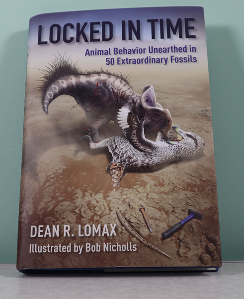
Dean Lomax is also the author of “Locked in Time”, a book which examines animal behaviour preserved in fifty exceptional fossil discoveries.
The front cover of the book “Locked in Time – Animal Behaviour Unearthed in 50 Extraordinary Fossils” by Dean Lomax with illustrations by Bob Nicholls. Picture credit: Everything Dinosaur.
Picture credit: Everything Dinosaur
Everything Dinosaur acknowledges the assistance of a media release from the University of Manchester in the compilation of this article.
The scientific paper: “A new giant theropod dinosaur track from the Middle Jurassic of the Cleveland Basin, Yorkshire, UK” by Hudson, J. G., Romano, M., Lomax, D. R., Taylor, R. and Woods, M. published in the Proceedings of the Yorkshire Geological Society.
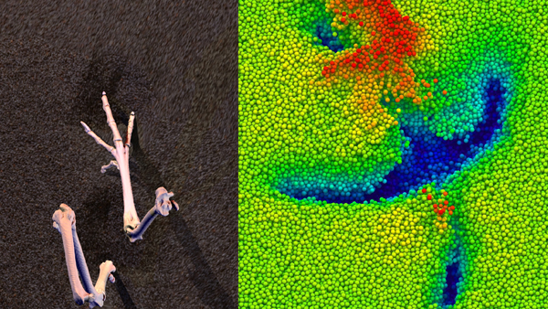
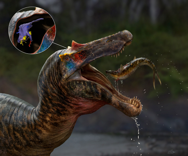
Researchers from the University of Southampton and Ohio University have recreated the brains and the inner ears of two early members of the Spinosauridae in a bid to better understand how these unusual theropods evolved as piscivores.
The spinosaurs Baryonyx walkeri and Ceratosuchops inferodios are the oldest members of the Spinosauridae family for which braincase material is known. When Baryonyx was formally named and described in 1986 (Charig and Milner), it helped revolutionise our understanding of these bizarre and enigmatic carnivorous dinosaurs. Ceratosuchops was scientifically described much more recently (2021). Several of the authors of the paper on Ceratosuchops participated in this study.
Having reconstructed the brains of these early spinosaurs, the researchers concluded that Baryonyx and Ceratosuchops brains were reminiscent of the brains of other theropods and lacked the specific adaptations and characteristics of later spinosaurs.
An artist’s impression of Ceratosuchops inferodios and the orientation of the endocast in the skull. Picture credit: Anthony Hutchings.
Spinosaurids – Not Your Usual Theropods
The Spinosauridae are considered unusual members of the theropod clade. Their evolutionary origins and their exact placement within the Theropoda remain uncertain. These dinosaurs are united by having a series of adaptations that indicate a more specialised predatory role within the ecosystem. They seem to have specialised in catching fish, evolving long snouts, nostrils placed further up the skull and crocodile-like jaws that were lined with large numbers of conical teeth. Adaptations that distinguish the Spinosauridae from other theropod dinosaurs such as the allosaurs, abelisaurids and tyrannosaurs, which seem to have been more generalist hypercarnivores.
Scanning the Braincase and Digitally Reconstructing the Brain
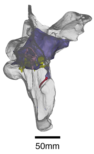
To help the researchers better understand the evolution of the spinosaur brain. The braincase fossils of Baryonyx and Ceratosuchops were scanned in high resolution. Sophisticated computer models of the brains, inner ears and related soft tissues of these two dinosaurs were created from these scans.
The digital reconstruction of spinosaur “grey matter” revealed that the olfactory bulbs, which process smells, weren’t particularly developed, and the ear was probably attuned to low frequency sounds. Those parts of the brain involved in keeping the head stable and the gaze fixed on prey were possibly less developed than they were in later, more specialised spinosaurs.
Three-dimensional reconstruction of the brain cavity and associated nerves and blood vessels within the braincase of the iconic British spinosaurid Baryonyx walkeri. Picture credit: WitmerLab/Chris Barker.
Commenting on the results, lead-author of the study, PhD student Chris Barker (University of Southampton), stated:
“Despite their unusual ecology, it seems the brains and senses of these early spinosaurs retained many aspects in common with other large-bodied theropods – there is no evidence that their semi-aquatic lifestyles are reflected in the way their brains are organised.”
Interpreting the Data
Although the fossil record of early spinosaurids is particularly poor, the researchers suggest that one interpretation of this brain study is that the theropod ancestors of spinosaurs already possessed brains and sensory adaptations that were suited to catching fish. Perhaps, as a way of avoiding direct competition with other large carnivores, the ancestral spinosaurids gradually spent more and more time hunting fish. Fish became an increasingly important part of the diet, a food resource not exploited to the same extent by other theropods. This led to the evolution of piscivorous adaptations such as longer jaws and conical teeth.
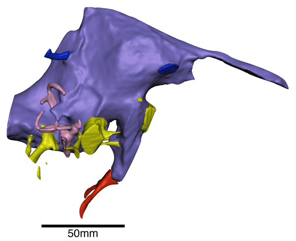
Three-dimensional reconstruction of the brain cavity (purple), cranial nerves (yellow), inner ear (pink) and blood vessels (red and blue) of the British spinosaurid Ceratosuchops inferodios. This predator likely had an unexceptional sense of smell and could hear low frequency sounds. Picture credit: Chris Barker.
British Spinosaurs Contributing to Palaeontology
Co-author of the study, published in the Journal of Anatomy, Dr Darren Naish (University of Southampton) stated:
“Because the skulls of all spinosaurs are so specialised for fish-catching, it’s surprising to see such ‘non-specialised’ brains. But the results are still significant. It’s exciting to get so much information on sensory abilities – on hearing, sense of smell, balance and so on – from British dinosaurs. Using cutting-edged technology, we basically obtained all the brain-related information we possibly could from these fossils.”
Learning More About the Spinosauridae
The non-destructive technique of using sophisticated computerised tomography (CT scans) in palaeontology is helping to change views and perceptions about the Dinosauria. Spinosaurs remain one of the most enigmatic and controversial families within the Theropoda. This new research mapping the brains and inner ears of early members of the Spinosauridae provides a valuable contribution to the on-going discussions about the evolutionary development of spinosaurids and their role as specialist piscivores in Early Cretaceous dinosaur dominated terrestrial communities.
Everything Dinosaur acknowledges the contribution of a press release from the University of Southampton in the compilation of this article.
The scientific paper: “Modified skulls but conservative brains? The palaeoneurology and endocranial anatomy of baryonychine dinosaurs (Theropoda: Spinosauridae)” by Chris Tijani Barker, Darren Naish, Jacob Trend, Lysanne Veerle Michels, Lawrence Witmer, Ryan Ridgley, Katy Rankin, Claire E. Clarkin, Philipp Schneider and Neil J. Gostling published in the Journal of Anatomy.
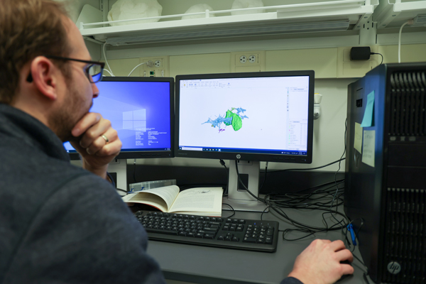
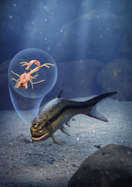
A team of international scientists including researchers from the University of Birmingham have published a paper on the brain and cranial nerves of fish that lived approximately 319 million years ago. The team’s findings are shedding light on vertebrate brain evolution.
The Late Carboniferous (early Pennsylvanian subperiod), fish fossil was discovered in a layer of soapstone adjacent to a coal seam at the Mountain Fourfoot coal mine in Lancashire and the specimen was first scientifically described in 1925. The fish, named Coccocephalus wildi, would have measured around 20 cm in length and it lived in what was an ancient estuary. It is only known from this single fossil and only the skull and jaws were recovered.
The fossilised skull of Coccocephalus wildi. The fish is facing to the right, with the jaws visible in the lower right portion of the fossil. The eye socket is the circular, bumpy feature above the jaws. Picture credit: Jeremy Marble, University of Michigan News.
Vertebrate Brain Evolution
Coccocephalus was a member of the Class Actinopterygii, also known as the ray-finned fishes. The skull fossil was sent on loan from Manchester Museum to the University of Michigan and subsequent CT scans of the skull revealed the surprising discovery of the intact brain and associated nerves.
Senior author Sam Giles, (University of Birmingham), commented:
“This unexpected find of a three-dimensionally preserved vertebrate brain gives us a startling insight into the neural anatomy of ray-finned fish. It tells us a more complicated pattern of brain evolution than suggested by living species alone, allowing us to better define how and when present day bony fishes evolved.”
University of Michigan palaeontologist Matt Friedman examines CT scan images of an exceptionally preserved, brain of the Late Carboniferous ray-finned fish Coccocephalus wildi. Picture credit: Jeremy Marble, University of Michigan News.
Rapidly Buried
When the fish died, it was probably buried rapidly in sediment containing very little oxygen. The lack of oxygen prevented the soft brain tissue from decaying. Whilst brain cases can reveal the shape and structure of vertebrate brains, this remarkable fossil preserved the brain tissue of a prehistoric fish.
Soft tissues such as the brain normally decay quickly and very rarely fossilise. But when this fish died, the soft tissues of its brain and cranial nerves were replaced during the fossilisation process with a dense mineral that preserved, in astonishing detail, their three-dimensional structure.
This discovery provides palaeontologists with a window into the evolution and development of the brains of ray-finned fishes, a highly successful group of back-boned animals estimated to represent more than fifty percent of all living vertebrate species.
Life reconstruction of the ray-finned fish Coccocephalus wildi showing location and shape of brain and cranial nerves. Picture credit: Márcio L. Castro.
A study of the jaws and teeth of C. wildi suggest that it was carnivorous, likely feeding on small invertebrates. The CT scans revealed that the brain had bilateral symmetry, like the brains of modern ray-finned fishes, but significantly, the brain of Coccocephalus folds inward, unlike in all living ray-finned fishes, in which the brain folds outward.
The fossil captures a time before a signature feature of ray-finned fish brains evolved, providing an indication of when this trait evolved.
Co-author of the paper, published in the journal “Nature”, Matt Friedman (University of Michigan) explained:
“An important conclusion is that these kinds of soft parts can be preserved, and they may be preserved in fossils that we’ve had for a long time—this is a fossil that’s been known for over 100 years.”
Everything Dinosaur acknowledges the assistance of a media release from the University of Birmingham in the compilation of this article.
The scientific paper: “Exceptional fossil preservation and evolution of the ray-finned fish brain” by Rodrigo T. Figueroa, Danielle Goodvin, Matthew A. Kolmann, Michael I. Coates, Abigail M. Caron, Matt Friedman and Sam Giles published in Nature.