Newly Described Multituberculate Mammal Provides Clues to Middle Ear Evolution
A team of international scientists from the Chinese Academy of Sciences, the American Museum of Natural History (New York) and the Beipiao Pterosaur Museum of China, have described a new species of multituberculate mammal that once roamed and climbed in the forests of north-eastern China during the Early Cretaceous. The small creature has been named Sinobaatar pani and its delicate middle ear bones have been preserved providing researchers with an opportunity to study the evolutionary development of hearing.
A Life Reconstruction of the Small Multituberculate Mammal S. pani
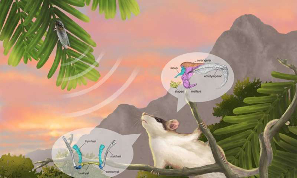
Picture credit: Institute of Vertebrate Palaeontology and Palaeoanthropology (IVPP)
How Do Mammals Hear?
The terrestrial placental mammal ear can be divided into three sections:
- Outer ear – collects and directs sound via the ear flap (pinna) and the outer ear canal which ends in the eardrum (tympanic membrane).
- Middle ear – which filters and amplifies sound waves directing them to the inner ear. The middle ear contains three, delicate and tiny bones (ossicles), the anvil, hammer and the stapes (incus, malleus and stirrup).
- Inner ear – consisting of the cochlea (organ for hearing) and the vestibular system (associated with balance).
The Inner Bones of a Model Mammal
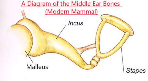
Picture credit: Everything Dinosaur
It is the inner ear bones that act as a bridge between the eardrum and the oval window which is the opening to inner ear. The cochlea, which is a hollow, spiral shaped bone transduces the sound waves into electrical signals (neural impulses), that are deciphered by the brain.
Sinobaatar pani and Middle Ear Evolution
Scientists believe that bones that were once part of the reptilian jaw slowly evolved into the three bones that are now found in the middle ear. A joint research team led by Dr Mao Fangyuan from the Institute of Vertebrate Palaeontology and Palaeoanthropology (IVPP) of the Chinese Academy of Sciences and Professor Meng Jin from the American Museum of Natural History were able to model the delicate middle ear bones of S. pani by using computerised tomography to access and view the fossils whilst they were still surrounded by matrix.
The powerful, rock-penetrating X-rays allowed the scientists to construct three-dimensional computer models of the malleus, incus and the stapes and to study their shape.
The images generated, permitted the comparing of the tiny inner ear bones of the Early Cretaceous multituberculate with the embryos of different types of living mammal (placental, marsupial and monotreme).
The Ancestral Phenotype of the Mammalian Middle Ear
Dr Mao explained that her colleagues were able to examine and assess the ancestral phenotype of the mammalian middle ear. The researchers recognised that the three main Mesozoic mammalian groups (multituberculates, eutriconodontans and symmetrodontans) share a similar middle ear structure between the incus and malleus, which they termed the “braced hinge joint”. Although they acknowledged that the middle ear may have evolved independently in several mammalian groups, they proposed that the braced hinge joint could represent a critical feature of the ancestral phenotype of the mammalian middle ear.
The Evolution of Mammalian Hearing
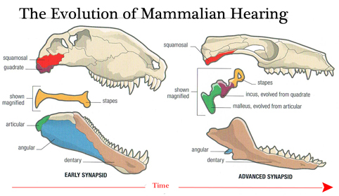
Dr Mao commented:
“There are two basic patterns of the middle ear in living mammals, represented by monotremes and therians [placentals and marsupials], respectively. In the former, the middle ear is characterised by an “abutting contact” between the incus and malleus, which is distinct from the one in therian mammals where the incus-malleus articulation is saddle-shaped.”
Sinobaatar pani Provides Clues
The abutting pattern in monotremes and the saddle-shaped joint in therians may well be derived from the braced hinge joint linking the incus and malleus as observed in Mesozoic mammals. At the very least, these fossil forms have narrowed the morphological gap between the middle ear of protomammals, formed by the postdentary bones lodged in the lower jaw, to the middle ear of extant mammals. The researchers proposed that the surangular bone, which is another postdentary bone in protomammals, persisted in Mesozoic mammals; its fate in living mammals remains uncertain.
Writing in the National Science Review, the researchers demonstrate that middle ear morphologies in Mesozoic mammals represent different evolutionary stages with Sinobaatar showing an advanced inner ear configuration. Furthermore, the evolutionary changes recorded in the Mesozoic mammals are largely consistent with the way the middle ear bones develop as living mammals grow, supporting the relationship between evolution and development.
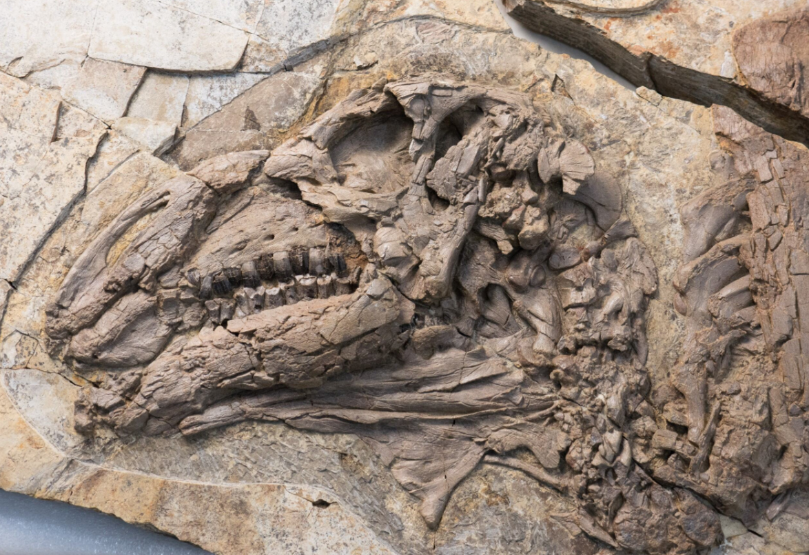
The Holotype of S. pani and Three-dimensional Skull and Teeth Images
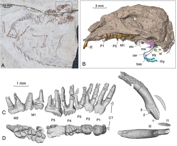
Picture credit: Institute of Vertebrate Palaeontology and Palaeoanthropology (IVPP)
Everything Dinosaur acknowledges the assistance of a press release from the Chinese Academy of Sciences in the compilation of this article.
The scientific paper: “Exploring ancestral phenotypes and evolutionary development of the mammalian middle ear based on Early Cretaceous Jehol mammals” by Fangyuan Mao, Cunyu Liu, Morgan Hill Chase, Andrew K Smith and Jin Meng published in National Science Review.
The Everything Dinosaur website: Everything Dinosaur.




Leave A Comment