Confusion over Dinosaur Colour – It’s an Inside Job
Internal Sources of Preserved Melanin Throw Doubt on Dinosaur Colour
One of the most exciting discoveries in the last two decades or so when it comes to the Dinosauria, was the recognition that fossilised, microscopic structures containing melanin (called melanosomes), could provide an indication of colour. The shape of the preserved melanosome when compared to the same type of structures found in living animals, gave scientists an insight into the potential colouration of long extinct creatures.
However, new research from a team of palaeontologists led by scientists from Bristol University and University College Cork (Ireland), has upset the colour scheme somewhat. They have discovered new sources of the pigment melanin such as in the liver, lungs and spleen preserved in fossils, this means how palaeontologists reconstruct the dinosaurs and other extinct prehistoric animals is going to have to be revisited.
Implications for the Dinosauria
This study has implications for the interpretation of colour within the Dinosauria.
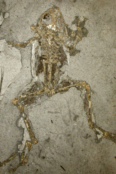
A Fossil Frog from the Miocene of Spain – Dark Areas in the Chest Cavity and Legs are Melanosomes
Picture credit: Museo Nacional de Ciencias Naturales, Madrid, (Spain)
Study Published in “Nature Communications”
Writing in the academic journal “Nature Communications”, the researchers looked at living and extinct amphibians. It is known that extant vertebrates have melanosomes within internal tissues, the scientists demonstrated that these internal melanosomes have a high fossilisation potential and can vastly outnumber those from the skin. This means that there could be a bias in fossils for preserved internal melanosomes thus “blurring” the picture when it comes to interpreting the colour of extinct animals.
Back to the Drawing Board? Do we Really Know the Colour of Extinct Creatures?
Picture credit: Everything Dinosaur
The picture (above) shows a brown Papo Giganotosaurus dinosaur model. Is this an accurate reflection of Dinosauria colouration?
To view the Papo dinosaur model range: Papo Dinosaur Models and Figures.
Powerful microscopes in conjunction with the chemical analysis of body tissues demonstrated that internal melanosomes are abundant. Lead author of the study, Dr Maria McNamara (University College Cork), stated:
“This means that these internal melanosomes could make up the majority of the melanosomes preserved in some fossils.”
However, all might not be lost as according to Dr McNamara, the shape and size of skin melanosomes are usually different from the melanosomes found in internal tissue. If this is the case, it might permit palaeontologist to refine their melanosome assessments, differentiating between internal and skin melanosome types. This could lead to more accurate depictions of extinct animals including dinosaurs.
Experiments with Decaying Frogs
Dr Paddy Orr (University College Dublin), along with Dr McNamara’s PhD student Valentina Rossi, also participated in the research, plotting the decay profiles of frogs in order to gain more information on how internal melanosomes can leak into other body parts during the fossilisation process.
Collaborator, Professor Mike Benton from the University of Bristol’s School of Earth Sciences, commented:
“Understanding the origin of melanosomes is crucial in the new studies of colour in dinosaurs and other extinct beasts.”
Slab and Counter Slab Splitting Can Influence the Picture
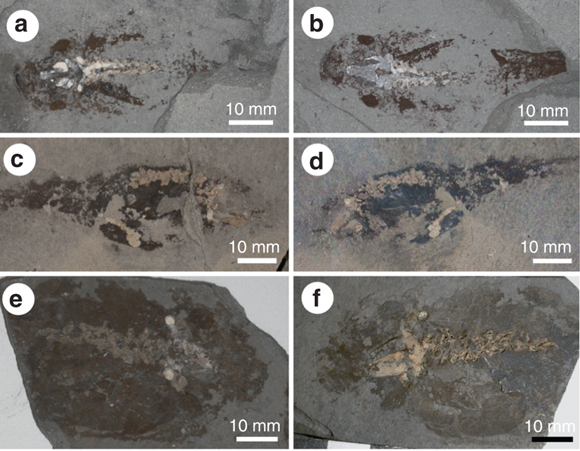
How a fossil is found in a slab or concretion can also influence any analysis of melanosomes. The researchers examined the distribution of soft tissues in the slab and counter slab of fossil amphibians. If the plane of splitting passes through the middle of the soft tissues, non-integumentary melanosomes can be exposed at the surface, producing nearly identical distributions of soft tissues in the slab and counter slab.
Distribution of Soft Tissues in the Slab and Counter Slab

Picture credit: Nature Communications
The photographs (above), labelled a to f, show near identical distributions of soft tissues in the slab and counter slab components of a fossil. However, if the fossil is split, not quite in the middle (not a medial split), then the slab and counter slab are likely to show different distributions of soft tissue components. Internal melanosomes may still be exposed at the surface of the fossil.
Uneven Distribution of Soft Tissue Between Slab and Counter Slab

Picture credit: Nature Communications
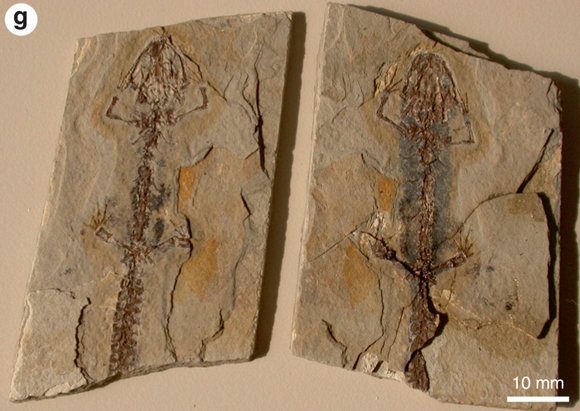
The picture (g), is the slab and counter slab of the prehistoric salamander Chelotriton from the Miocene of Spain. The part of the fossil on the right shows more black staining, the remnants of soft tissues preserved in the fossil. The plain of splitting of this fossil is not precisely medial, therefore leading to one side of the fossil showing greater staining than its counterpart.
A spokesperson from Everything Dinosaur commented:
“This newly published research, rather than thwarting attempts to reconstruct dinosaurs and other prehistoric animals, might actually lead to a refining of the illustration process, allowing palaeontologists and the palaeoartists that work closely with them to create more accurate reconstructions.”
Everything Dinosaur acknowledges the assistance of a media release from the University of Bristol in the compilation of this article.
Visit the Everything Dinosaur website: Everything Dinosaur.


